
Figure 3.
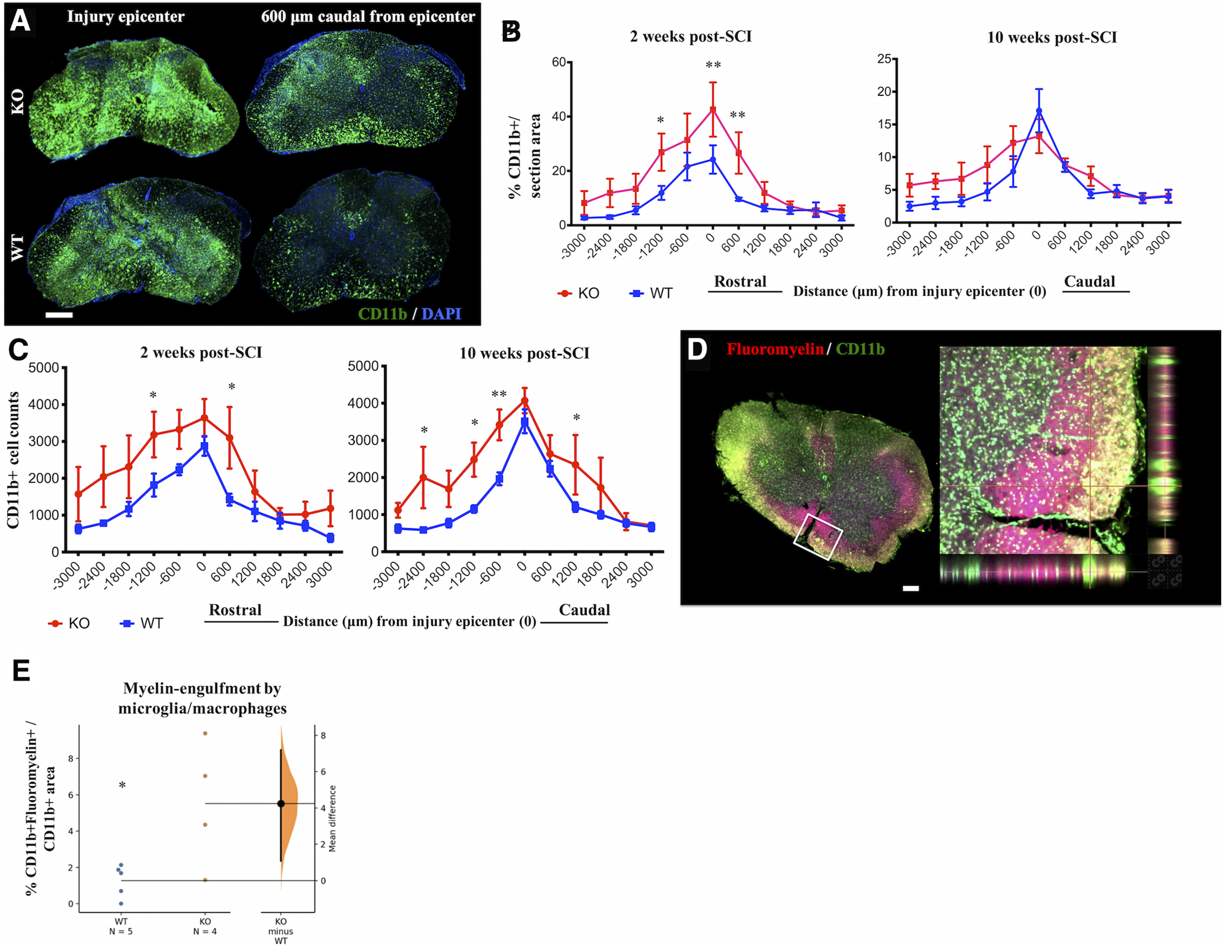
IgM-KO mice have more microglia/macrophages than WT controls. A, Representative spinal cord sections stained for the microglia/macrophage markers CD11b and DAPI at two weeks post-SCI. Quantification of % CD11b+ immunostained areas/total area (B) and CD11b+ cell counts (C) rostro-caudally from the injury epicenter, at 2 and 10 weeks of injury. D, Representative spinal cord section stained for myelin (fluoromyelin) and microglia/macrophages (CD11b) at two weeks post-SCI. Inset indicates area of co-localized CD11b and fluoromyelin signal. Myelin-engulfing macrophages appear yellow because of colocalization of green (CD11b+ signal) and red (fluoromyelin) signals in the single layer confocal image. E, Gardner–Altman estimation plot showing quantification of % area of co-localized CD11b+ and fluoromyelin+ signal/total section area. Each dot indicates mean as averaged from 3 sections/animal, where sections were taken at distances −1200, −600, and +600 μm from the injury epicenter. Scale bar: 100 μm, N = 4–5 animals/group (B, E), 5 animals/group (C). Error bars indicate SEM; *p < 0.05, **p < 0.01. See Table 1 for detailed statistical results in B, C and Table 2 for statistical results in E.