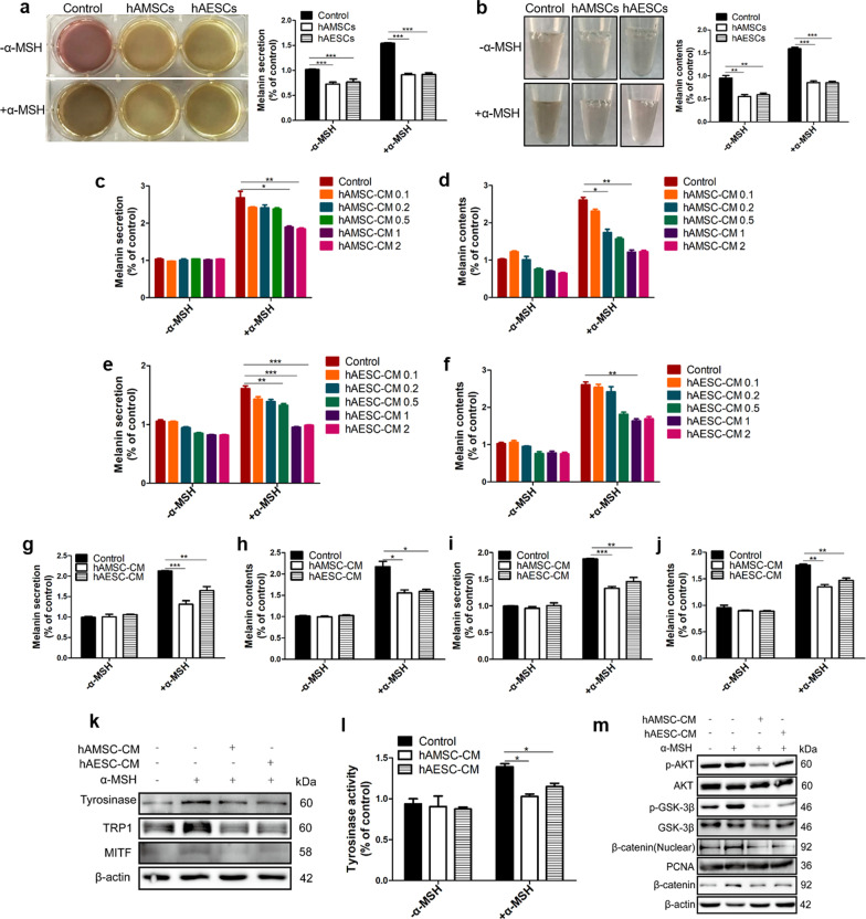
Fig. 2.
hAMSC-CM or hAESC-CM inhibits α-MSH-induced melanogenesis in B16F10 cells. hAMSCs or hAESCs were co-cultured with B16F10 for 2 days and melanin pigments were quantified by measuring absorbance values of cell supernatant (a) and cell precipitation suspension (b). B16F10 cells were stimulated with α-MSH and then treated with various concentrations of hAMSC-CM or hAESC-CM (concentrations from 2 × 10⁴ to 2 × 10⁶ cells/mL, and hAMSC-CM 1 or hAESC-CM 1 indicates the concentration of 2 × 10⁵ cells/mL) for 48 h, the cell supernatants (c, e) and cell pellets (d, f) were obtained to determine anti-melanogenesis effect of hAMSC-CM (c, d) or hAESC-CM (e, f). B16F10 cells were pre-treated with α-MSH (200 nM) for 6 h and then further co-incubated with hAMSC-CM or hAESC-CM for 48 h. The cell supernatants (g) and cell pellets (h) were obtained for determining the therapeutic effects of hAMSC-CM or hAESC-CM as described in the materials and methods. B16F10 cells were pre-treated with hAMSC-CM or hAESC-CM for 6 h, then α-MSH was added to further incubate for 48 h, and the cell supernatants (i) and cell pellets (j) were obtained for determining protective effects of hAMSC-CM or hAESC-CM. The effects of hAMSC-CM or hAESC-CM on the protein expression levels of melanogenesis-related protein (tyrosinase, TRP1 and MITF) were analyzed by western blotting analysis (k). The effects of hAMSC-CM or hAESC-CM on tyrosinase enzyme activity were confirmed by treatment with L-DOPA and detection of tyrosinase at 475 nm (l). The phospho-AKT, phospho-Gsk3β, β-catenin and β-catenin (nuclear) were determined by western blot analysis in B16F10 cells with α-MSH stimulation (m). Data are presented as mean ± SD. The experiments were repeated three times independently and the data of one representative experiment was shown. *p < 0.05; **p < 0.01, ***p < 0.001

