
Fig 2. MmPEG10 protein and mRNA is secreted in vesicles by cells in vitro.
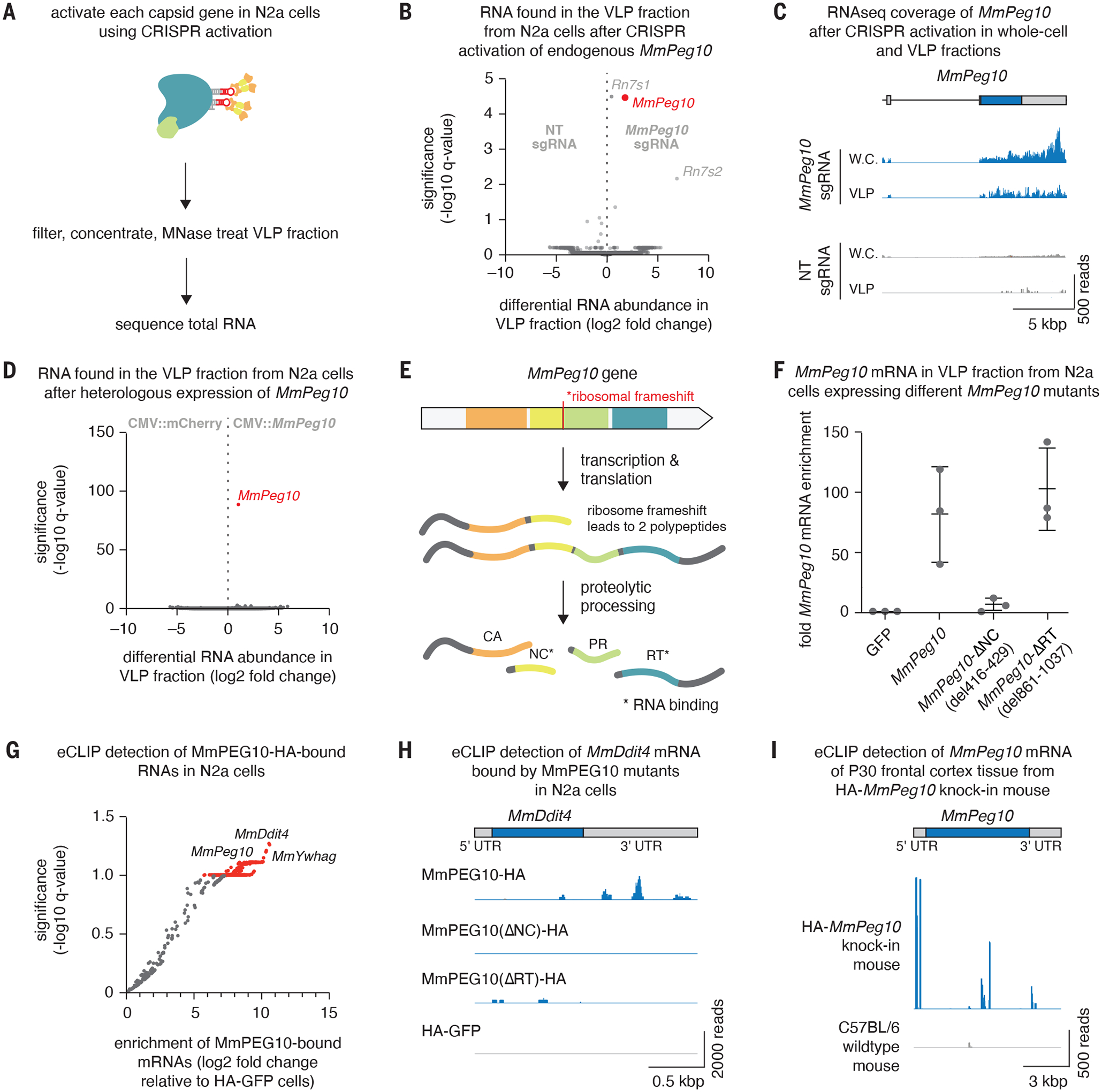
A. Method for identifying nucleic acids that are secreted in the VLP fraction upon gene activation of CA-domain containing proteins.
B. Differential RNA abundance and significance in the VLP fraction from N2a cells after CRISPR activation of endogenous MmPeg10.
C. Alignment of sequencing reads showing sequencing coverage of the MmPeg10 mRNA from (B).
D. Differential RNA abundance and significance in the VLP fraction from N2a cells after heterologous transfection of MmPeg10. n=3 replicates.
E. The four domains of MmPEG10 are translated into two isoforms. These are self-processed by the PEG10 protease into separate domains, of which the NC and RT bind RNA.
F. Fold enrichment of MmPeg10 mRNA compared to GFP in the VLP fraction from N2a cells transfected with wildtype MmPeg10 or deletions of the predicted nucleocapsid (ΔNC) and reverse transcriptase (ΔRT) domains.
G. Log2 fold change and significance of bound RNAs from eCLIP data comparing HA-GFP to WT MmPEG10-HA.
H. Representative sequencing alignment histogram of the MmDdit4 locus generated from eCLIP of N2a cells transfected with wildtype or mutant MmPeg10.
I. Representative sequencing alignment histogram of the MmPeg10 locus generated from eCLIP data of n = 3 HA-PEG10 and n = 3 untagged animals.