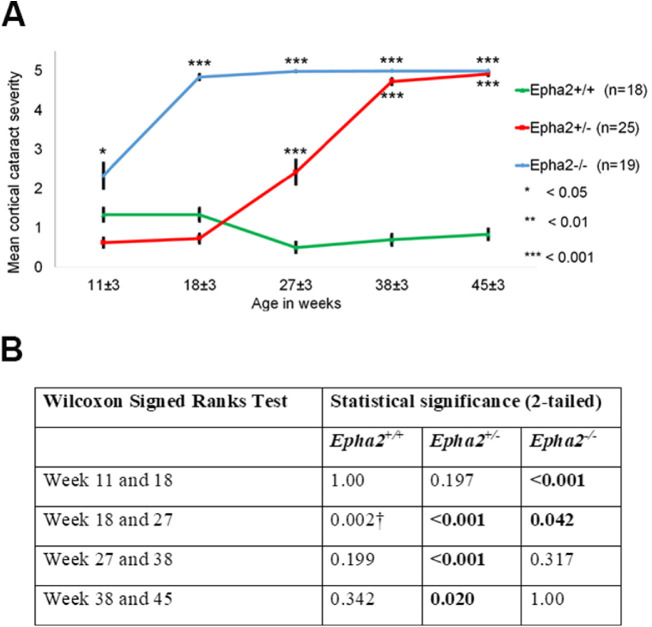
Figure 2.
Progression of anterior cortical cataract in Epha2-knockout mice.Epha2+/+, Epha2+/−, and Epha2−/− mice on C57BL/6J background were monitored for cataract development from 11 ± 3 weeks to 45 ± 3 weeks of age. (A) Mean cortical cataract grade in each genotype of mice at each time point has been graphically represented. Epha2−/− mice developed severe cortical cataract at 18 ± 3 weeks of age and Epha2+/− mice at 38 ± 3 weeks of age. Epha2+/+ mice presented with very mild lens opacity up to 45 ± 3 weeks of age. The Mann Whitney U test P values from comparison with Epha2+/+ mice are indicated. Error bars represent standard error of the mean. After an animal exhibited a cataract grade of 4 or 5, the same grade was attributed to subsequent time points for data analysis, assuming no further increase in cataract severity occurred. Missing data at the first time point was substituted with data from the second time point assuming lesser cataract severity at the first than second time point. (B) The table shows Wilcoxon Signed Ranks test P values of comparison between time points indicating the effect of age on cataract progression in each genotype of mice. † Indicates likely artifact of anesthetic-induced cataract.

