Figure 4.
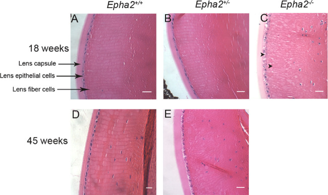
Histological analysis showing disruption of lens architecture in Epha2-knockout mouse lenses. Representative images of hematoxylin and eosin-stained sections of lenses of 18-week-old Epha2+/+ (A), Epha2+/− (B), and Epha2−/− (C) mice and 45-week-old Epha2+/+ (D) and Epha2+/− (E) mice on C57BL/6J background showing nuclei (blue) and cytoplasm (pink) of lens epithelial and fiber cells as well as lens capsule. Disruption of fiber cell arrangement in the lens cortex can be seen in 18-week-old Epha2−/−, C, and 45-week-old Epha2+/−, E, mouse lenses as opposed to arrangement in meridional rows in Epha2+/+, A and D, and 18-week-old Epha2+/− lenses, B. Arrowheads show presence of vacuoles in the lens cortex and lens epithelial cells. Nucleated fiber cells are visible in those sections closer to the lens equator. Scale-bars 20 µm.

