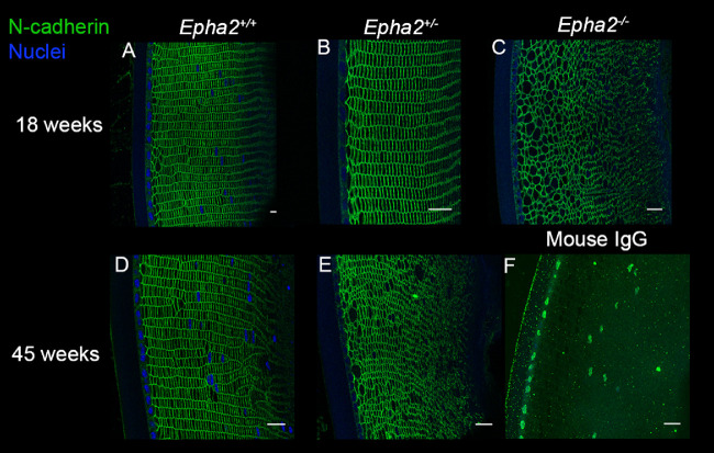
Figure 5.
N-cadherin immunolabeling showing disorganization of cellular architecture in Epha2-knockout mouse lenses. Representative confocal microscopy images of sections of lenses of 18-week-old Epha2+/+ (A), Epha2+/− (B), and Epha2−/− (C), and 45-week-old Epha2+/+ (D) and Epha2+/− (E) mice on C57BL/6J background immunolabeled with mouse anti-N-cadherin antibody (green) and DAPI (blue) to label the nuclei are shown. N-cadherin labeling delineated the cell boundary and showed well packed fiber cells arranged in meridional rows in 18, A, and 45-week-old Epha2+/+, D, and 18-week-old Epha2+/−, B, lenses. In contrast, it showed larger and disorganized fiber cells in 18-week-old Epha2−/−, C, and 45-week-old Epha2+/−, E, lenses. Sections probed with mouse IgG (negative control) revealed little or no non-specific labeling (F). Scale-bars 20 µm.

