Figure 7.
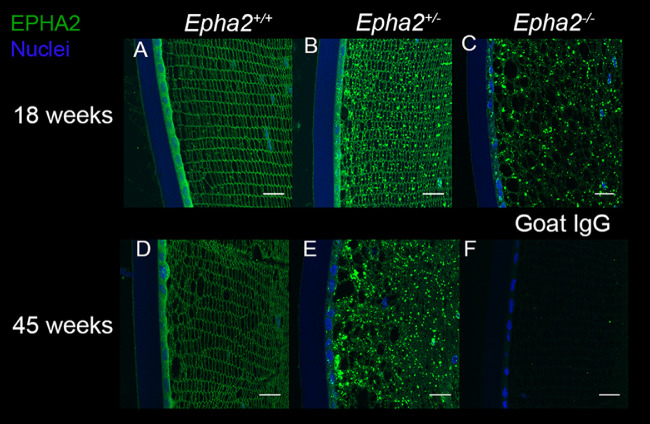
Accumulation of mutant EPHA2 protein in Epha2-knockout mouse lenses detected by immunolabeling. Representative confocal microscopy images of lens sections of 18-week-old Epha2+/+ (A), Epha2+/− (B), and Epha2−/− (C) and 45-week-old Epha2+/+ (D) and Epha2+/− (E) mice on C57BL/6J background were immunolabeled with anti-mouse EPHA2 antibody (green) and DAPI (blue) to stain the nuclei. The lenses of 18, A, and 45-week-old Epha2+/+, D, showed the EPHA2 protein in lens epithelial cells and at the lens fiber cell periphery. Eighteen-week-old lenses of Epha2+/− mice, B, in addition to the protein in lens epithelium and lens fiber cell periphery, exhibited its presence in a granular pattern in cells. Lenses of 45-week-old Epha2+/−, E, and 18-week-old Epha2−/−, C, mice showed presence of the protein only in granular pattern likely indicating accumulation of the partial EPHA2-β-galactosidase fusion protein in lens cells. Similar labeling was absent in negative control sections incubated with goat IgG (F). Scale-bars 20 µm.

