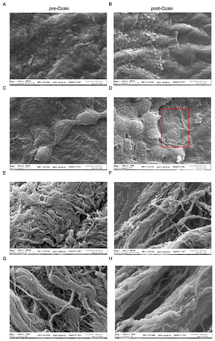
Figure 2.
SEM morphological analysis of pre-Ozaki and post-Ozaki samples. (A,C) Representative images of the serosa layer in the pre-Ozaki pericardium show a continuous layer of mesothelial cells, whose surface is covered by short and abundant microvilli. (B,D) This layer appears interrupted in some areas (red box) in the post-Ozaki pericardium. (E,G) Representative images of the surface of the fibrosa layer in the pre-Ozaki pericardium display overlapping wavy collagen and elastic fibers. (F,H) These fibers assume a more parallel and compact arrangement in the post-Ozaki pericardium. Representative images at 1500× (A,B,E,F) and 7000× (C,D,G,H) magnifications.

