
FIGURE 4.
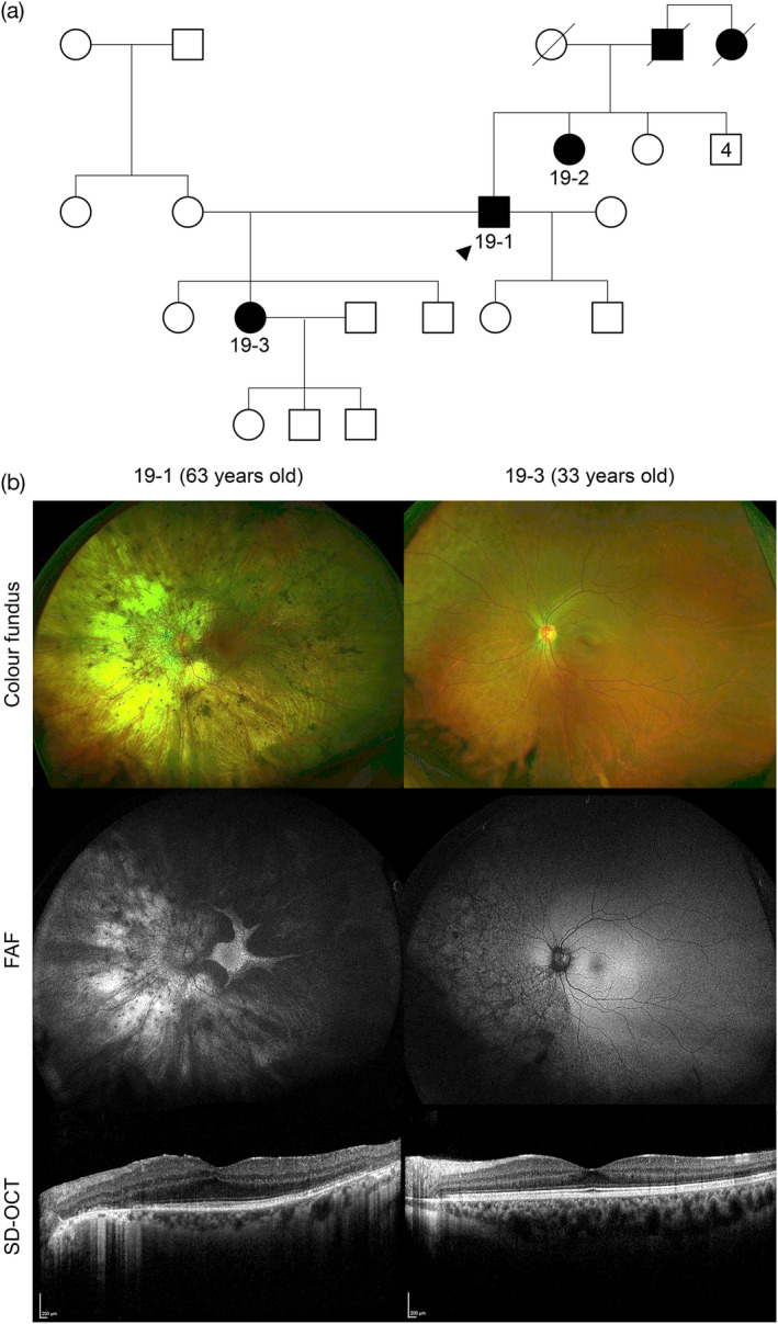
Family 19 has a novel heterozygous multi‐exon (1–8) duplication of TEAD1, associated with Sveinsson chorioretinal atrophy. (a) Family tree highlighting the autosomal dominant inheritance, no consanguinity, with proband (19‐1, arrowhead), his affected sister (19‐2), daughter (19‐3) and deceased father and paternal aunt. (b) The clinical phenotype of the left eye is shown (right eye had symmetrical findings) of the proband 19‐1 aged 63 years and his affected daughter (19‐3) aged 33 years. In proband 19‐1, there is an extensive widespread chorioretinal atrophy, more marked on the nasal side and peripapillary region, with a preserved central macular retinal island on the UWF color fundus imaging. FAF imaging shows a well delineated hyperautofluorescent retinal island with scalloped edges, the superotemporal retina has hypoautofluorescence. SD‐OCT with a horizontal line scan through the fovea shows relatively well‐preserved ellipsoid zone with clear edges of outer retinal layer disruption and loss. In patient 19‐3, there are RPE changes extending from inferonasal peripapillary margin to the far nasal retina, corresponding to changes in the FAF, which shows scalloped hypoautofluorescence throughout this area. SD‐OCT shows a healthy macula with intact ellipsoid zone