
FIGURE 4.
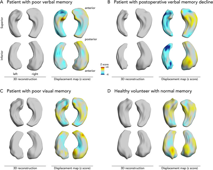
Three‐dimensional (3D) reconstructions of hippocampi in example cases. Displayed are 3D reconstructions of the hippocampal surface. The z scores of inward (blue, ie, atrophy) or outward (yellow/red, ie, hypertrophy) displacements from a mean normal hippocampal template are projected onto these surfaces. (A) Hippocampi of a 41‐year‐old female with left temporal lobe epilepsy with poor presurgical verbal memory (25 points) and slightly impaired visual memory (30 points) but no verbal memory decline after surgery (35 points). She had left hippocampal atrophy mainly affecting the left hippocampal head. (B) Hippocampi of a 63‐year‐old female with left temporal lobe epilepsy with slightly impaired presurgical verbal (41 points) and visual (26 points) memory and verbal memory decline following surgery (24 points). She had left hippocampal atrophy affecting the left hippocampal head and tail. (C) Hippocampi of a 65‐year‐old female with right temporal lobe epilepsy with normal presurgical verbal memory (44 points) and poor visual memory (12 points) but no verbal or visual memory decline after surgery (49 and 10 points, respectively). Despite normal hippocampal volumes, there was localized atrophy that affected the inferomedial hippocampal surface mainly on the right. (D) Hippocampi of a 30‐year‐old healthy male with good verbal and visual memory performance (73 and 43 points, respectively).