
Figure 5. Ihh is required for teriparatide stimulation of MMPs and progenies.
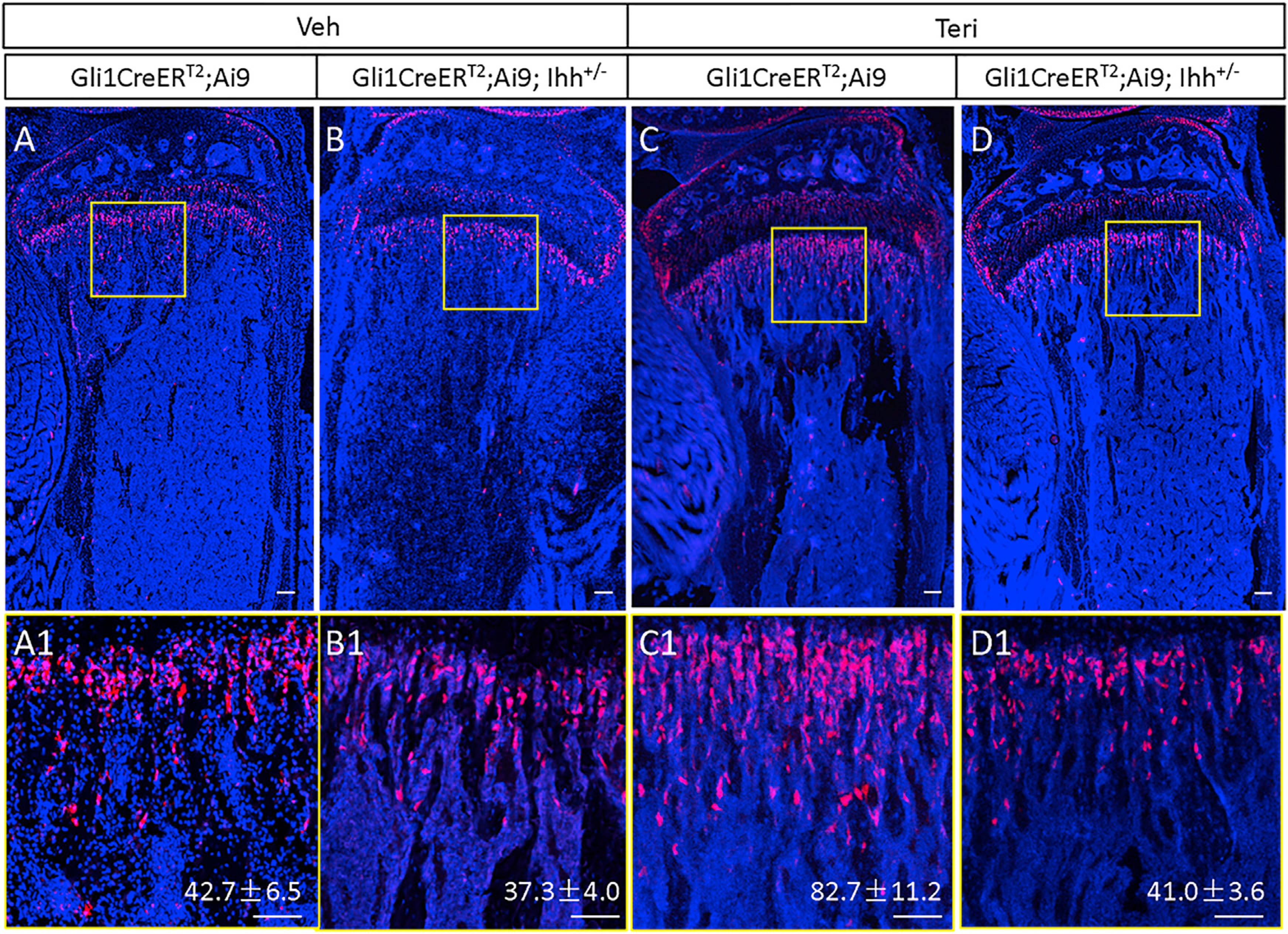
Mice of indicated genotypes were injected at one month of age with teriparatide or vehicle for three days and then TAM for three days.
(A–D) Confocal images of longitudinal sections through the proximal tibia.
(A1–D1) Boxed regions in (A)–(D) shown at higher magnification. Red, tdTomato+ MMPs and progenies; blue, DAPI nuclear staining. Scale bar: 100 μm. tdTomato+ cells were counted in a primary spongiosa area of 0.09 mm2 (300 × 300 μm) immediately below the growth plate. Quantification indicates mean cell number ± SD, n = 3. One section per mouse was quantified.