Table 1.
Common in vitro mechanical force stimulation methods and their major studied outcomes
| Mechanical force | Description | Major outcomes | Benefits | Drawbacks |
|---|---|---|---|---|
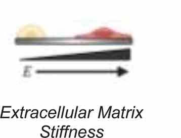 |
Stiffening or softening of extracellular matrix to induce mechanical responses similar to that of native tissue [124,134,240,241] | • Focal adhesion activation • Actin cytoskeleton polymerization • Nuclear stiffening • Cell differentiation • Chromatin organization |
• Replicates to native tissue mechanics • No additional apparatus required to induce mechanical signals • No additional apparatus required to induce mechanical signals |
• Can have uneven stiffness profiles across surfaces • Harder to image live or fixed cells |
 |
Restricting cell shape through physical impediments or shape of adherent surface [32–35,242] | • Cytoskeleton & nucleus shape • Cell differentiation • Chromatin organization |
• Easy to manufacture and implement • Isolates function of cell shape in cellular functions • Can image live or fixed cells |
• Low cell density • Partial homology to tissue environment |
 |
Mimicry of fluid shear stress forces found in vasculature systems [31,112–115,243,244] | • Cell and nucleus orientation • Cytoskeleton remodeling |
• High homology to vasculature forces • Easy to mimic human pathologies |
• Requires use of specially designed bioreactors • Fluid force can be non- uniform between experiment sets |
 Strain Strain
|
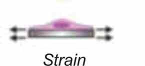
Stretching of adherent substrate to produce dynamic or static strain forces [6,7,13–17,37,52,56,100,127] | • Actin cytoskeleton • Cell differentiation • Cell proliferation • Focal adhesion signaling • Nuclear signaling and structure • Chromatin organization |
• Easy to use • Induces strong regulation of differentiation and stimulation of the actin cytoskeleton |
• Requires expensive strain application machinery • Limited by size of specialized cell culture plates |
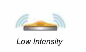 |
Low magnitude strain induced by low amplitude, high-frequency vibration [19,37,53,55,56,100] | • Focal adhesions signaling • Cell differentiation • Cell proliferation • Nuclear signaling and structure |
• Similar homology to muscle-induced vibration forces observed in native tissue • Can be utilized in cell culture, tissues, and mammalian models |
• Requires custom-made bioreactors • Requires long-term exposure to mechanical signals • Less potent mechanical signal compared to strain and fluid shear |
 |
Probing of individual cells and nuclei with rounded-tip atomic force microscopy [100,145,147,169,245] | • Measure Cell and nuclear stiffness • Force induced translocation of mechanically sensitive biomolecules |
• Provides high resolution stiffness measurement of cells and nuclei • Targeted mechanical activation of mechanosensitive signaling pathways |
• Require expensive equipment Challenging to provide provide population-based measurements • Hard to determine if measuring proper target versus non-desired targets |
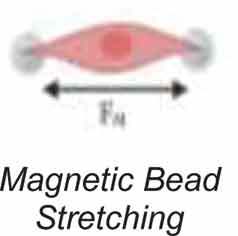 |
Use of magnetic beads to induce physical strain on individual cells [136,246–248] | • Force induced translocation of mechanically sensitive biomolecules • Nuclei mechanoresponse •Actin cytoskeleton remodeling • Chromatin |
• Allows for targeted strain on an individual cell level • Can induce targeted chromatin structure changes |
• Does not provide population-based measurements • Requires use of special equipment |
