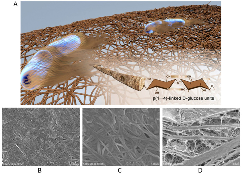
Figure 2.
(A) A representative model image of a 3D nanofibrous network of BNC as secreted by A. xylinus bacteria (including detailed hydroxyl functional groups of the highlighted single nanofibril). Reproduced with permission from [29]. Copyright 2016 Elsevier. SEM images of BNC surface at different magnifications as (B) 10.0 kx and (C) 50.0 kx. (D) cross-section of BNC (5.0 kx). Reproduced from [28].

