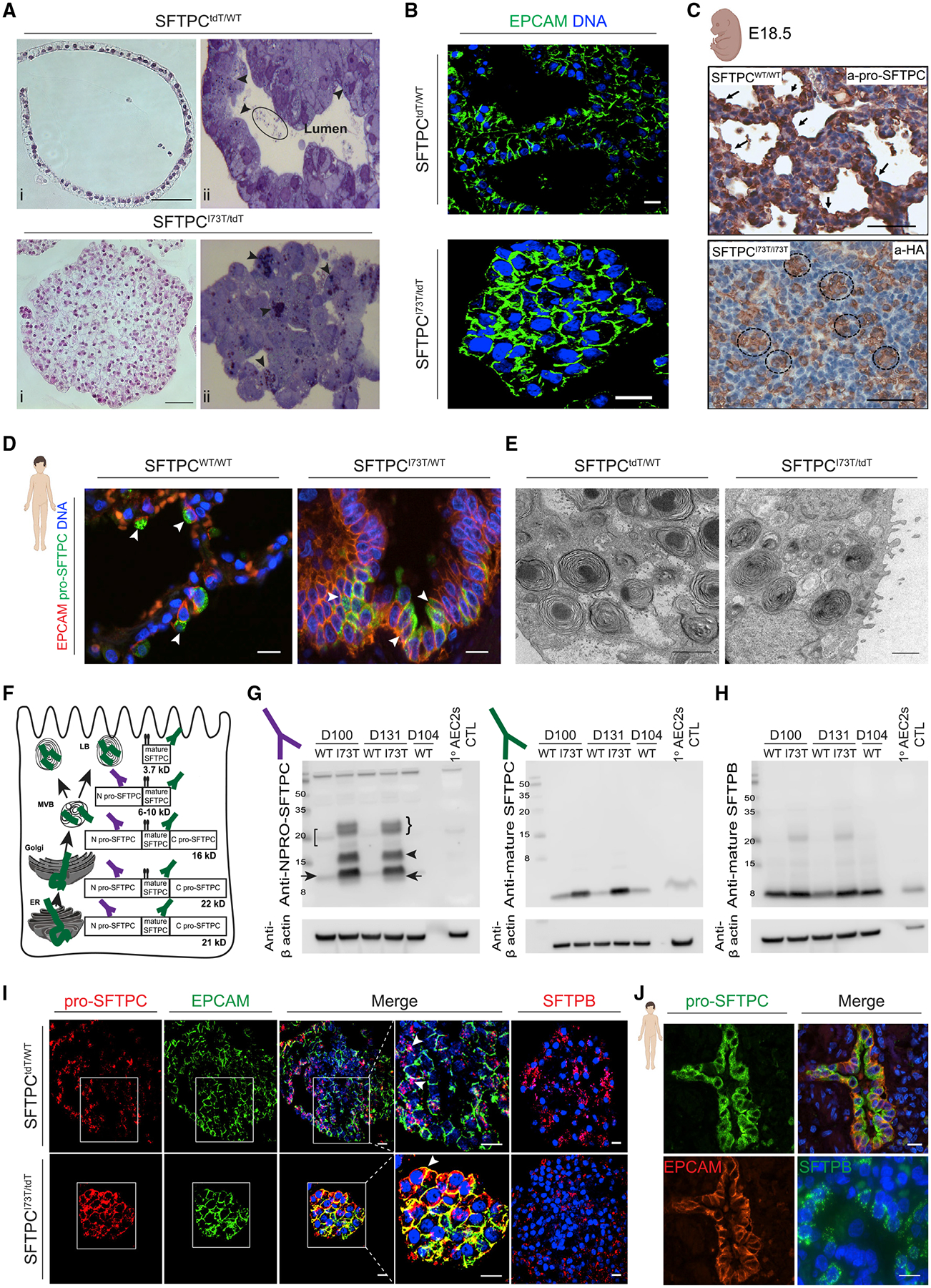
Figure 3. SFTPCI73T/tdT iAEC2s demonstrate distinct cellular morphology and misprocess and mistraffick pro-SFTPC similarly to in vivo SFTPCI73T-expressing AEC2s.

(A) Representative H&E staining of formalin fixed and paraffin embedded sections (i, scale bars, 50 μm) and toluidine blue staining of plastic sections (ii) of SFTPCtdT/WT and SFTPCI73T/tdT iAEC2s. Arrowheads indicate lamellar body-like inclusions; eclipse indicates intraluminal inclusions.
(B) Representative confocal immunofluorescence microscopy of SFTPCtdT/WT and SFTPCI73T/tdT iAEC2s for EPCAM (green) and DNA (Hoechst, blue). Scale bars, 10 μm.
(C) Representative staining of E18.5 CFlp-SFTPCI73T/I73T and CFlp-SFTPCWT/WT embryos for HA-tagged SFTPC. Scale bars, 70 μm.
(D) Representative confocal immunofluorescence microscopy of distal sections of SPC2 donor and healthy donor lung explants stained for EPCAM (red), pro-SFTPC (green), and DNA (Hoechst, blue). Scale bars, 10 μm.
(E) Representative TEM images of SFTPCtdT/WT and SFTPCI73T/tdT iAEC2s depicting lamellar bodies. Scale bars, 1 μm.
(F) Schematic representing the cellular compartments in which pro-SFTPC processing into mature SFTPC occurs. ER, endoplasmic reticulum; MVB, multi-vesicular body; LB, lamellar body.
(G and H) Representative western blots of SFTPCtdT/WT and SFTPCI73T/tdT iAEC2 lysates at the indicated time points were compared to freshly isolated primary human AEC2s lysates for pro-SFTPC (NPRO-SFTPC) (G, left panel), mature SFTPC (G, right panel), and mature SFTPB (H) with β actin as a loading control.
(I) Representative confocal immunofluorescence microscopy of SFTPCtdT/WT and SFTPCI73T/tdT iAEC2s for pro-SFTPC (red; with zoom), EPCAM (green; with zoom), SFTPB (red), and DNA (Hoechst, blue). Scale bars, 10 μm.
(J) Representative confocal immunofluorescence microscopy of distal sections of SPC2 donor and healthy donor lung explants for pro-SFTPC (green), EPCAM (red), SFTPB (green), and DNA (Hoechst, blue). Scale bars, 10 μm.
