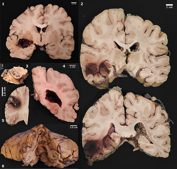
Fig 1. Gross pathology of macroscopic hemorrhages.
1. Hemorrhage in the putamen; 2. Extensive hemorrhage in temporal lobe; 3. Punctate hemorrhages in cerebral peduncles; 4. Lateral ventricle filled by blood; 5. Focal hemorrhage in cerebellar cortex; 6. Punctate hemorrhages in middle cerebellar peduncles.

