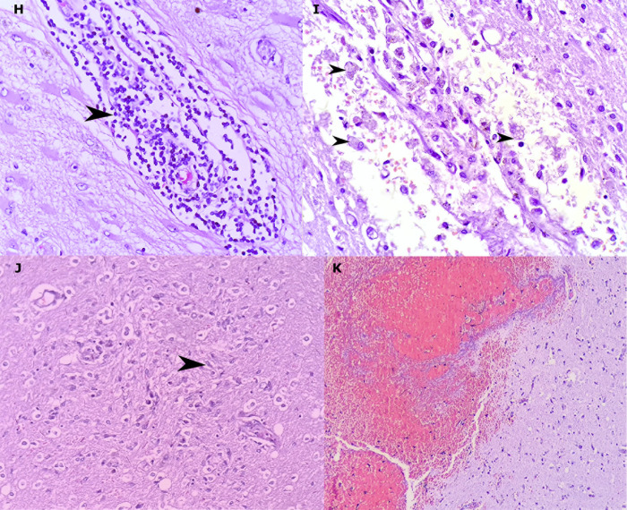
Fig 4. Inflammatory cells and reaction processes in the examined brains.
Lymphocytes (arrowhead) forming bulky perivascular cuffing (H). Macrophages (arrowhead), red cell diapedesis and edema around blood vessels (I). Microglial nodule (microglia cell, arrowhead) (J). Occurrence of intraparenchymal hemorrhagic phenomenon without association with inflammatory process (K). (hematoxylin and eosin. H: 400x, I: 200x, K: 200x).

