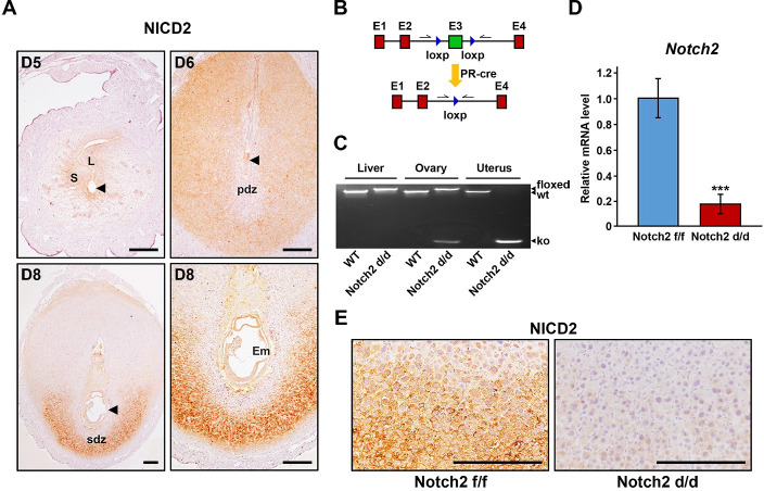
Fig 1. Notch2 was efficiently deleted in the uterus.
(A) The spatiotemporal expression pattern of NICD2 in the uterus during early pregnancy was revealed by immunohistochemistry. Arrowheads indicate the developing embryo. L, luminal epithelium; S, stroma; pdz, primary decidual zone; sdz, secondary decidual zone; Em, embryo. Scale bar: 100μm. (B) The scheme shows the strategy of uterine-conditional Notch2 ablation. Arrows indicate primers used for the examination of knockout efficiency. (C) Genotyping analysis was carried out to validate knockout efficiency at DNA level in the liver, ovary and uterus. (D) Knockout efficiency at mRNA level in the uterus was further confirmed by QRT-PCR. Data are presented as mean±SEM. (E) Immunohistochemistry staining of NICD2 revealed that Notch2 was efficiently ablated at protein level in the uterus. Scale bar: 100μm. ***p<0.001.

