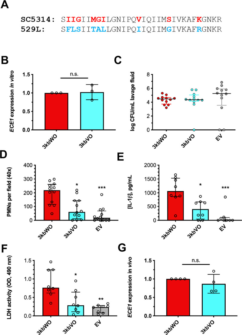
Fig 2. Isolate 529L harbors an alternative candidalysin allele.

(A) Diagram depicting conserved (black), SC5314-like (red), 529L-like (blue) amino acids. (B) In vitro ECE1 expression levels were assessed by qRT-PCR 4 h after strains were transferred to RPMI-1640 and normalized to ACT1 and 3kbWO using the ΔΔCt method (mean ± SD). Statistical significance was assessed using a Student’s t-test. Mice (n = 12 per group) were challenged with 3kbWO, 3kbVO, and empty vector strains and vaginal lavage performed at d 3 post-infection. Lavage fluids were assessed for (C) CFU by microbiological plating (median ± IQR), (D) PMN recruitment by microscopy (median ± IQR), (E) IL-1β by ELISA (median ± IQR), and (F) tissue damage by LDH assay (median ± IQR). Statistical significance was assessed using Kruskal-Wallis and Dunn’s post-test. (G) ECE1 expression (mean ± SD) was assessed in vivo by qRT-PCR as fold-change of ECE1 over ACT1 and normalized to 3kbWO using the ΔΔCt method. Significance was assessed using a Student’s t-test. All in vitro experiments were conducted in biological triplicate. *, p < 0.05, ** p < 0.01, *** p < 0.001.
