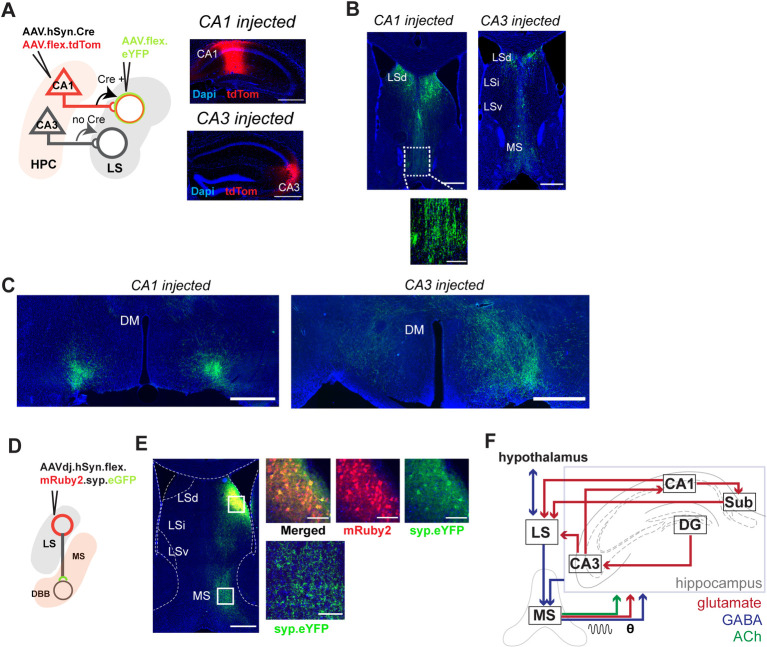
Fig 6. LS cells receiving hippocampal inputs project directly to hypothalamus and MS.
(A) Diagram of dual-viral injection strategy for anterograde transsynaptic tracing. The diagram is based on dorsal CA1 targeted injection; dCA3 is also used for injections, together with coronal sections showing primary injection sites in dorsal CA1 (top) or dorsal CA3 (bottom). Red: tdTom expression; blue: DAPI, with zoomed images showing tdTom-positive cell bodies are predominantly located in the pyramidal layer (bottom). For additional images of spread of injection, see S19 Fig. (B) eYFP-positive cell bodies at the anterior dorsal LS and fibers at the level of the MS following dCA1 injection (left) and dCA3 injection (right). Inset left: eYFP-positive fibers at the level of MS. (C) eYFP-positive axons are seen bilaterally at the level of the LH following dCA1 injection (left) and dCA3 injection (right). (D) Injection strategy for Cre-dependent AAV. Synaptag mediated tracing in the LS. (E) Coronal section of dorsal LS, with synaptophysin-bound eYFP at the MS. Top right: zoomed images showing transduction at the injection site. Bottom right: eYFP-positive fibers. (F) Schematic with proposed connections of the LS within the hippocampal network. Scale bars: B, left and right: 800 μm. C, left and right: 500 μm. The underlying data can be found in S1 Data. DBB, diagonal band of Broca; DG, dentate gyrus; DM, dorsomedial hypothalamic nucleus; HPC, hippocampus; LH, lateral hypothalamus; LS, lateral septum; LSd, dorsal lateral septum; LSi, intermediate lateral septum; LSv, ventral lateral septum; MS, medial septum.

