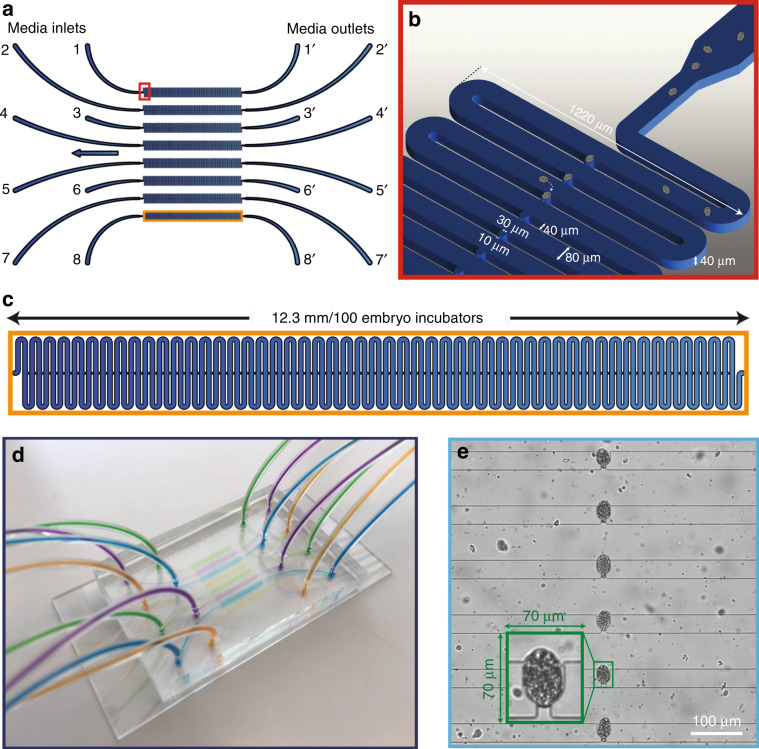
Fig. 1. Details of the high-throughput microfluidic platform used for automated phenotyping of C. elegans embryos.
a Schematic representation of the microfluidic chip with eight multiplexed lanes with one end connected to the media inlet and the other to the outlet. b Zoomed-in image of the entrance of a microfluidic lane with the feature sizes marked. c Image of a single lane that consists of 100 embryo incubators within 12.3 mm. d A picture of the PDMS chip (35 × 50 mm) filled with dye solutions and bonded onto a standard glass microscope slide (38 mm × 75 mm). e An image of six embryo incubators visualized during time-lapse imaging; a single incubator containing an embryo (in green) is displayed that was utilized by the automated script for phenotyping

