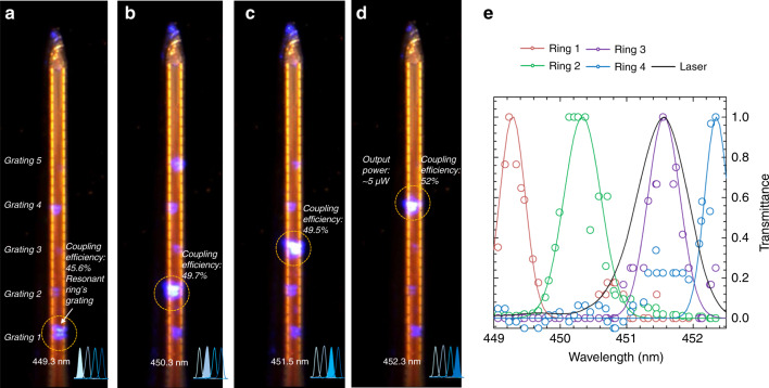
Fig. 5. In vitro probe optical characterization.
Optical microscope image of the tip of the assembled neural probe after turning on the laser at the first four ring resonance wavelengths: a 449.3 nm, b 450.3 nm, c 451.5 nm, and d 452.3 nm. The number of each grating is reported in (a) and is the same in (b–d). For each image, we report the resonant grating coupling efficiency. All the gratings in the image are visible through oversaturation for graphical purposes. The scale bar is 50 µm. e Normalized experimental ring transmittances for different ring resonators (colored circles, with the Gaussian fits reported as solid lines in corresponding colors) and laser spectrum (solid black line)

