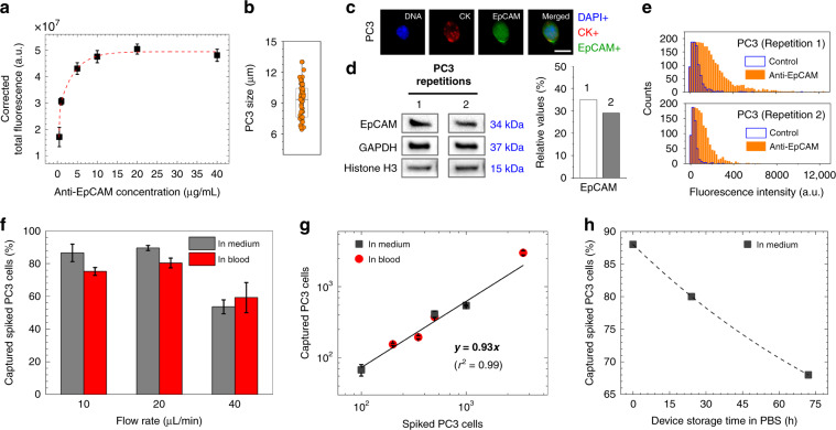
Fig. 3. Device and cell characterization.
a Variations in anti-EpCAM antibody adsorption capacity (n = 3) suggested that 10 μg/mL antibody concentration saturates maleimide-activated glass substrate (after BMPS deposition) of the microfluidic device. b Sizes of PC3 cells ranged from ~6 to 13 μm in diameter with an average size of ~9 μm. c Fluorescent image of a PC3 cell (DAPI+/CK+/EpCAM+), showing a cytoplasmic-to-nuclear area ratio of ~2.32. The scale bar is 10 μm. d Surface EpCAM protein levels in pelleted PC3 cells, as revealed by western blotting analysis, were ~32% overexpressed compared to the combined levels of GAPDH (control) and histone H3 (control). e Surface EpCAM protein levels in PC3 cells, as revealed by flow cytometry, verified that >75% of cell surface EpCAM binds to fluorophore-conjugated anti-EpCAM antibodies. f One milliliter of culture medium or blood sample was used to evaluate the capture of 1000 spiked in PC3 cells at 10, 20, and 40 μL/min flow rates. Among the tested flow rates, a 20 μL/min flow rate resulted in capture of ~90% of cells when spiked into culture medium (n = 4) and ~80% of cells when spiked into healthy blood (n = 3). g The spiked number of PC3 cells in culture medium (1000, 500, and 100 cells/mL, n = 3) and blood (3500, 500, 350, and 200 cells/mL, n = 3) showed good linear correlation with the captured number of cells. h Storing anti-EpCAM antibody-activated microfluidic devices for 3 days in PBS at 4 °C resulted in an ~20% reduction in the capture efficiency of PC3 cells spiked in culture medium. Error bars represent the mean ± S.D

