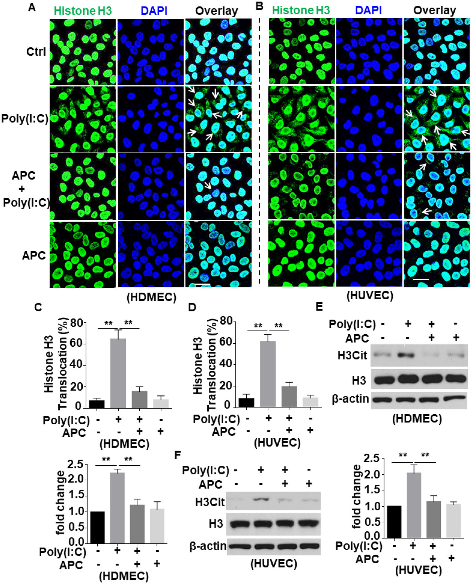
Figure 2. APC inhibits poly(I:C)-induced citrullination and extranuclear translocation of histone H3 in endothelial cells.

(A and B) HDMECs (A) or HUVECs (B) were pretreated with APC (20 nM for 3h) followed by stimulation with poly(I:C) (10 μg/mL for 1h) (APC remained in the media after addition of poly(I:C)). Cells were fixed and permeabilized followed by staining for histone H3 with mouse anti-histone H3 antibody and Alexa Fluor 488-conjugated goat anti-mouse IgG. The nucleus was stained with DAPI. Immunofluorescence images were taken by confocal microscopy. Arrows indicate extranuclear translocation of histone H3. (C and D) The quantification of poly(I:C)-mediated translocated cells from the nucleus to the extranuclear space for HDMECs (A) and HUVECs (B). (E and F) HDMECs (C) or HUVECs (D) were pretreated with APC (20 nM for 3h) followed by stimulation with poly(I:C) (10 μg/mL for 1h). Cell lysates were immunoblotted and probed with an antibody specific for unmodified or citrullinated histone H3 (H3Cit). β-actin was used as a loading control. Scale bar: 20 μm. Results are shown as means ± standard error from at least 3 independent experiments. **p < 0.01. Ctrl, control.
