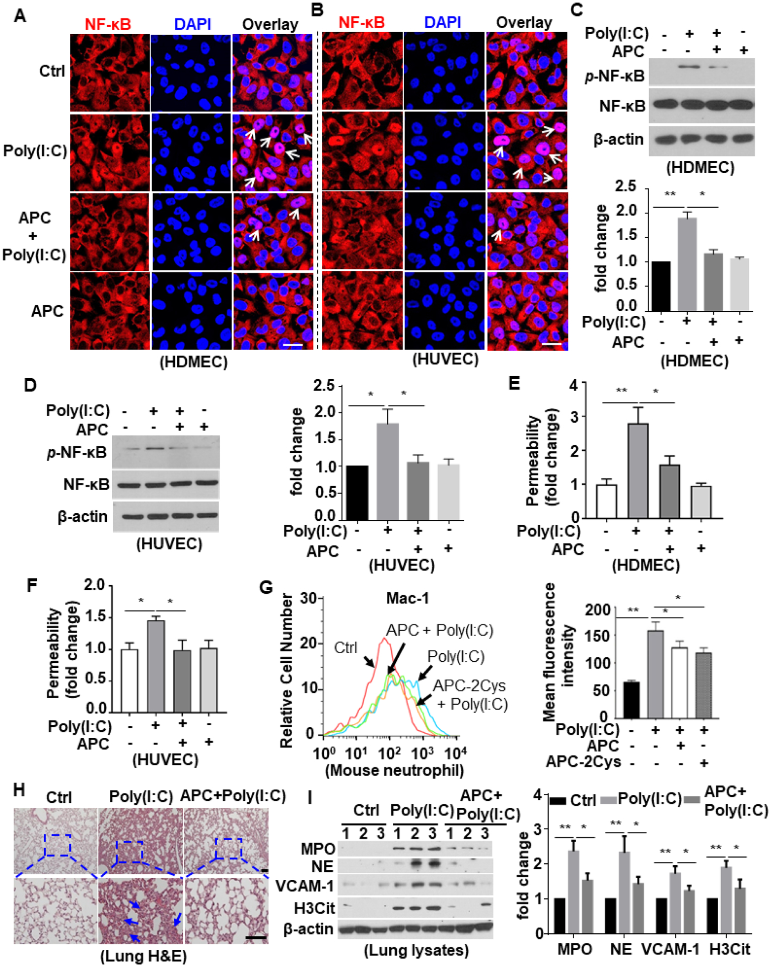
Figure 8. APC inhibits poly(I:C)-induced proinflammatory signaling.

(A and B) HDMECs (A) or HUVECs (B) were pretreated with APC (20 nM for 3h) followed by stimulation with poly(I:C) (10 μg/mL for 1h) (APC remained in the media after addition of poly(I:C)). Cells were then fixed, permeabilized and NF-κB p65 was stained with rabbit anti-NF-κB antibody and Alexa Fluor 555-conjugated goat anti-rabbit IgG. The nucleus was stained with DAPI. Immunofluorescence images were taken by confocal microscopy. Arrows indicate nuclear translocation of NF-κB. (C and D) HDMECs (C) or HUVECs (D) were pretreated with APC (20 nM for 3h) followed by stimulation with poly(I:C) (10 μg/mL for 1h). Cell lysates were immunoblotted and probed with an antibody specific for phosphorylated NF-κB (p-NF-κB). β-actin was used as a loading control. (E and F) HDMECs (E) or HUVECs (F) were pretreated with APC (20 nM for 3h) followed by stimulation with poly(I:C) (10 μg/mL for 4h). The amount of Evans blue dye that leaked into the lower chamber in the Trans-well assay plates was measured. (G) Mice were injected i.p. with APC or the signaling-selective mutant of APC (APC-2Cys) 1h prior to poly(I:C) i.p. injection for 3h and blood was collected and stained for Ly6G-FITC & Mac-1-PE. The cell surface expression of Mac-1 in Ly6G-FITC-positive neutrophil population was measured by flow cytometry. Representative data were obtained from 3–5 mice per group (n = 3–5). The mean fluorescence intensity was quantified. (H and I) Mice were pretreated with APC for 1h followed by i.p. injection with poly(I:C) for 3h, and lung tissue was collected and processed for histological analyses. Paraffin-embedded sections of lung tissue were stained with H&E (H). Representative images were obtained from 5 mice per group (n = 5). The inset boxes from each group are magnified. The arrows indicate inflammatory foci. The lung tissue was harvested for lysis. Tissue lysates were immunoblotted for myeloperoxidase (MPO), neutrophil elastase (NE), VCAM-1, citrullinated H3 (H3Cit) and β-actin. The relative expression levels of these proteins were quantified (I). Scale bar: 20 μm (A and B), 200 μm (H). All experiments were repeated at least three times. Results are shown as mean ± standard error. *p < 0.05, **p < 0.01.
