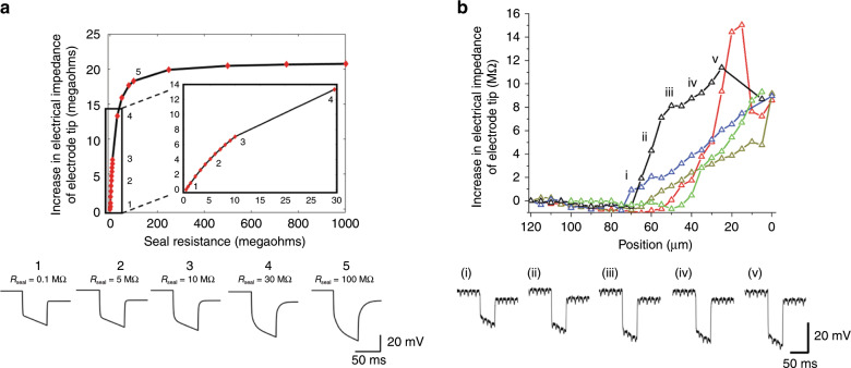
Fig. 4. Electrical impedance of the tip of GP microelectrode as it approaches a neuron in the isolated abdominal ganglion.
a The decreasing distance between electrode and neuronal membrane was simulated by increasing the seal resistance, Rseal. For each Rseal, voltage response of the GP microelectrode to a 1-nA current pulse was predicted and the electrical impedance of the electrode tip was measured using the algorithm in Fig. 3e. The voltage responses predicted by the model for five increasing values of Rseal are shown below. b Experimental measurements of change in electrical impedance of the tip of the GP microelectrodes with increase in proximity to neurons (n = 5 neurons). The increase in electrical impedance began 45–70 μm before successful cell penetration of the five neurons. The recorded voltage responses of an electrode at five different positions of the electrode with respect to a neuron are shown in the lower trace.

