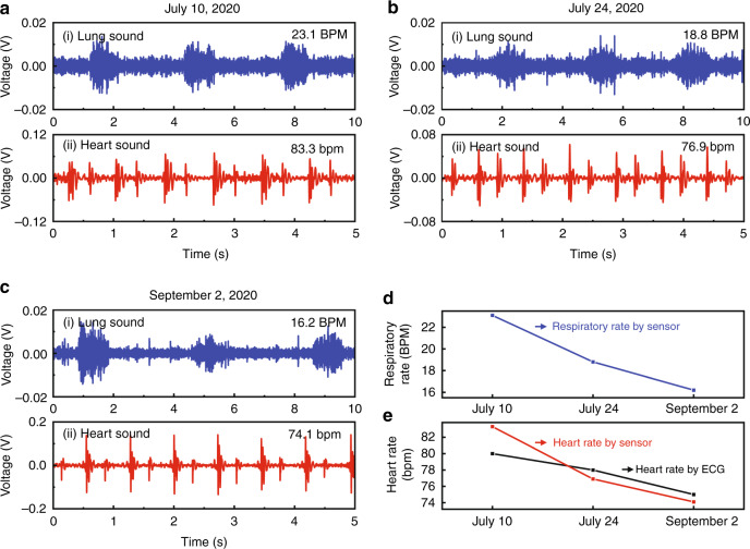
Fig. 7. Time evolution of lung and heart states of a discharged pneumonia patient.
a, b, c Sound sensor monitoring of the patient (#23) on July 10 (a), July 24 (b), and September 02, 2020 (c). ai, bi, ci Lung sounds recorded by our sensor; aii, bii, cii Heart sounds detected by our sensor. d Time evolution of lung sounds of the patient detected by sensor monitoring. e Time evolution of heart sounds of the patient detected by our sound sensor monitoring (red curve) compared with ECG data (black curve)

