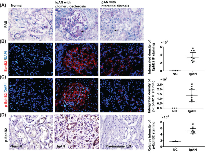
Figure 2. EphB2 expression increases in human kidney biopsy tissue with CKD compared with normal subjects.
Kidney biopsy sections from normal part of kidney (NC, n=3) and the IgAN patients (Oxford pathological type T = 1 [38]) (n=7) from Nanjing Medical University were stained with PAS (A), EphB2 (red) (B), and phosphor-EphB2 (p-EphB2) (red) (C). Magnification, ×200. DAPI-stained DNA (blue) served as nuclei staining control for IF staining (IF staining was carried out as described for mouse tissue). Use of the human material was approved by the local ethical review board. Magnification ×200. Arrows indicated fibrosis in glomerulus and tubulointerstitial area. (D) Immunochemistry staining for p-EphB2 (stained in brown) (using the same anti-pEphB2 antibody as the primary antibody as used for IF staining and the peroxidase-conjugated anti-rabbit IgG (Jackson ImmunoResearch Laboratories Inc.) as the secondary antibody and positive staining was developed by using 3,3′-diaminobenzidine) was further carried out on 3-µm sections of paraffin-embedded these IgAN kidney biopsy tissues and was counterstained with Hematoxylin. As a negative control, the primary antibody was replaced by pre-immune IgG and no specific staining was noted. Magnification ×400. Relative integrated pixel density of the staining of the EphB2 or p-EphB2 specially in renal tubulointerstitial area, as quantified by ImageJ, was shown on the right of each staining photos. *P<0.05 vs. normal kidney (NC).

