Figure 1.
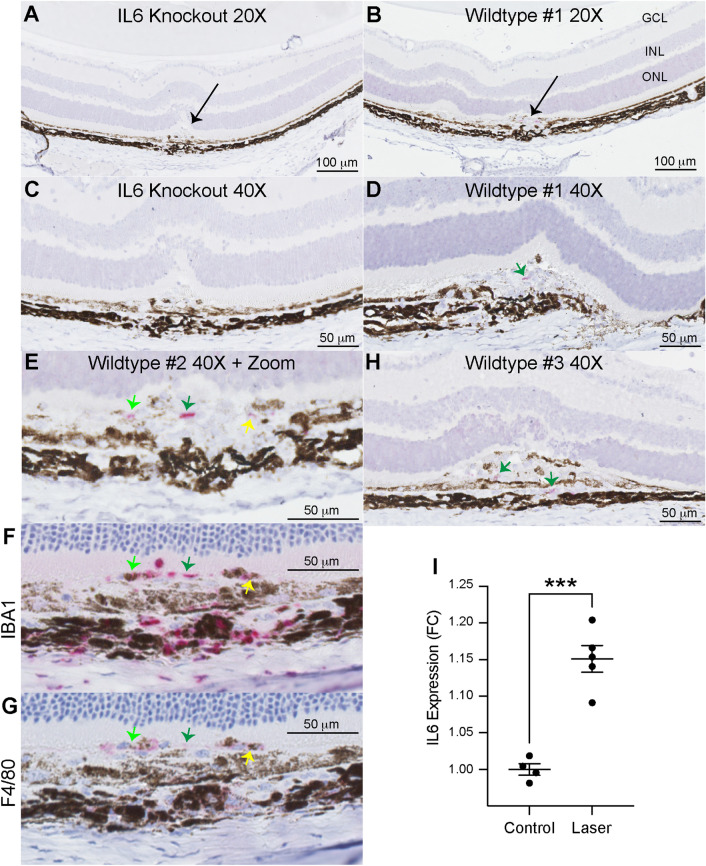
IL6 expression is increased by laser and expressed at the laser injury site. 20X magnification of paraffin-embedded sections stained for hematoxylin and IL6 (pink, RNAscope) from Il6−/− (A) and wildtype (B) mice. Arrows indicate laser lesion. 40X magnification of sections stained for hematoxylin and IL6 (pink, RNAscope) from Il6−/− (C) and wildtype (D, H) mice. Green arrows indicate IL6 + cells. 40 magnification with digital zoom of IL6 (E), IBA1 (F), and F4/80 (G) in serial sections from a single lesion. Light green, dark green, and yellow arrows indicate IL6 + cells co-staining for IBA1 and F4/80 in serial sections. ELISA measurements (I) of IL6 expression demonstrate a 1.15-fold (*** = p < 0.001, N = 4–5 per group) increase after laser injury.

