Figure 4.
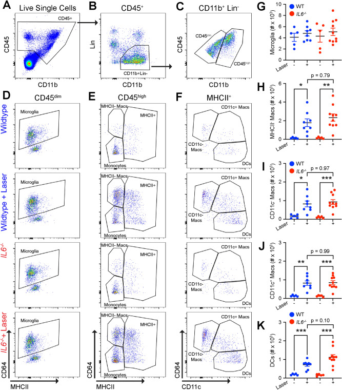
Wildtype and Il6−/− mice demonstrate similarly increased macrophage numbers. (A) CD45+ cells were identified from live and singlet cells. (B) Lineage (Lin) gate was used to exclude neutrophils (Ly6G), eosinophils (SiglecF), NK cells (NK1.1), T cells (CD4, CD8), and B cells (B220), and gate forward CD11b+Lin− mononuclear phagocytes. (C) Quantitative CD45 expression was used to separate CD45dim from CD45high cells. (D) Microglia were defined as CD45dimCD64+MHCIIlow from wildtype and Il6−/− mice both with and without laser. (E): MHCII− macrophages (macs) were delineated as CD45highCD64+MHCII−, and CD45highMHCII+ cells were gated forward. (F): CD11c− and CD11c+ macrophages were identified by CD64+MHCII+CD11c− and CD64+MHCII+CD11c+, respectively. Dendritic cells (DC) were designated as CD64−MHCII+CD11c+. (G) Microglia were not changed by genotype or laser. (H–J): MHCII−, CD11c−, and CD11c+ macrophages were equally increased by laser in wildtype (blue) and Il6−/− (red) mice (* = p < 0.05, ** = p < 0.01, *** = p < 0.001, N = 5–9 per group). (K) Dendritic cells were also increased by laser in both wildtype and Il6−/− mice.

