Figure 6.
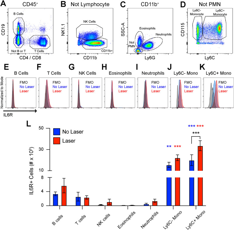
Monocytes express the IL6R in peripheral blood. (A) B cells were defined as CD19+, T cells were defined as CD4+ or CD8+, and non-lymphocytes (not B or T) were gated forward. (B) NK cells were delineated as NK1.1+ and CD11b+NK1.1− cells were gated forward. (C) Neutrophils were identified as Ly6G+SSCmed, eosinophils were found to be SSChighLy6G−, and non-granulocytes (Not PMN) were gated forward. (D) Monocytes were defined as CD115+Ly6C− or CD115+Ly6C+. (E–K) Representative frequency histograms for IL6R expression from the fluorescence minus one (FMO) control, untreated (No Laser, Blue), and lasered (Laser, Red) mice in B cells (E), T cells (F), NK cells (G), Eosinophils (H), Neutrophils (I), Ly6C− monocytes (J), and Ly6C+ monocytes (K). L: All cells other than monocytes were < 5% IL6R+. At steady state, Ly6C− and Ly6C+ monocytes expressed more IL6R than all other groups (** = p < 0.01, *** = p < 0.001 vs all other groups [Blue vs No Laser], N = 5–6 per group). After laser injury, more IL6R+Ly6C− and IL6R+Ly6C+ monocytes detected compared to all other groups (*** = p < 0.001 vs all other groups [Red vs Laser], N = 5–6 per group). The number of IL6R+Ly6C+ monocytes were increased by laser treatment (*** = p < 0.001, Black: No Laser vs Laser, N = 5–6 per group).

