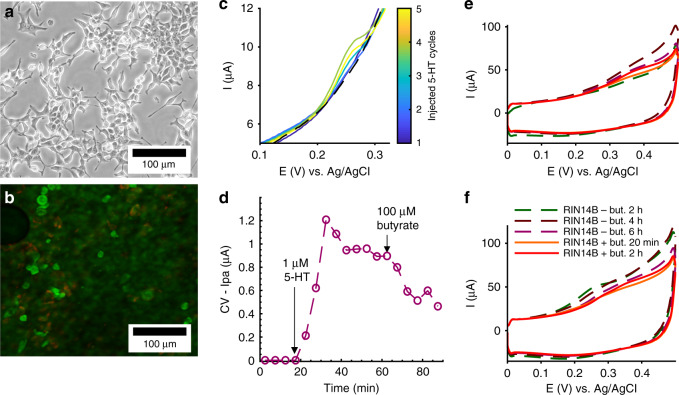
Fig. 8. Poor membrane attachment of RIN14B cells is a limiting factor for 5-HT detection from cells cultured on our electrode-integrated membrane.
a, b Optical micrographs of RIN14B cells. a Bright-field image of RIN14B on a T75 polystyrene flask. b Confocal fluorescence microscopy of live/dead stained RIN14B grown on an electrode-integrated cell culture membrane-coated with collagen. Live cells: green (Syto9), dead cells: red (propidium iodide). The cell morphology and density were analyzed. c, d RIN14B cells cultured on a Au-CNT (2 µL) membrane electrode coated with collagen were monitored over the course of molecular treatment, where c shows CV curves and d shows Ipa values measured from those curves. c CV curves show baseline (black—dashed) and exogenously injected 5-HT (color bar). d Time course of Ipa values, where arrows denote time of injections: spiking with 1 µM 5-HT for calibration, and stimulation with 100 µM butyrate. A 5min accumulation time was used between CV cycles. e, f RIN14B cells cultured on Au-CNT (12.5 µL) membrane electrodes, e with collagen and f without collagen, were monitored with a 2 h accumulation time between CV cycles. Dashed lines denote baseline measurements taken at t = 2, 4, and 6h. Solid lines denote measurements taken at t = 20 min and 2 h after the addition of 100 µM butyrate

