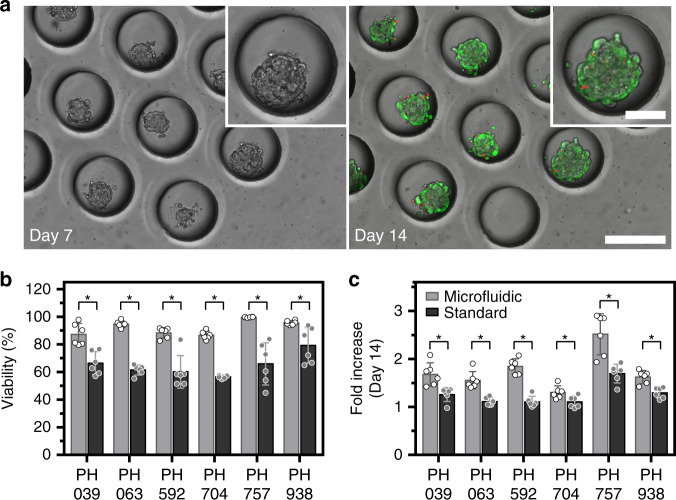
Fig. 2. Cultivation of OC spheroids in a single chamber microfluidic device.
a Micrographs showing microwell areas from a microfluidic device with spheroids after 7 and 14 days of culture (scale bar = 250 µm). The insert shows a zoomed-in image of a single microwell/spheroid (scale bar = 100 µm). The right image shows live/dead staining at day 14 (live (green), dead (red fluorescence)). b Viability of spheroids from the six PDX lines after 14 days of culture were tested in this study to compare microfluidic (gray bars) versus standard (black bars) culture methods. Live/dead staining was used for quantification (p < 0.05, n = 6). c Increase in spheroid diameter was observed after 14 days of culture (p < 0.05, n = 6)

