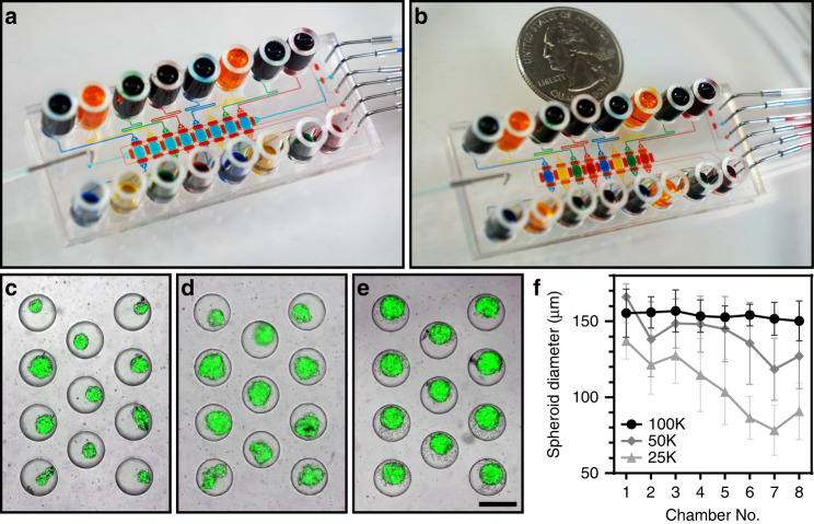
Fig. 5. A multichamber microfluidic device.
a, b Pictures showing a microfluidic device in serial perfusion (a) and parallel testing (b) modes. In serial perfusion, the device is configured to connect culture chambers serially. All culture chambers in (a) are filled with blue dye. In parallel perfusion, the device is reconfigured to sequester individual culture compartments and infuse them with dyes of different colors (blue, yellow, green, and red). c–e Images showing spheroids formed after seeding 25,000 (c), 50,000 (d), and 100,000 (e) cells into the device. Scale bar: 300 µm. f A graph showing spheroid diameter as a function of inoculation concentration for the 8 chambers of the device. Chamber 1 is closest to the inlet used for cell seeding, and chamber 8 is furthest. Spheroids were measured 24 h after seeding (n = 8)

