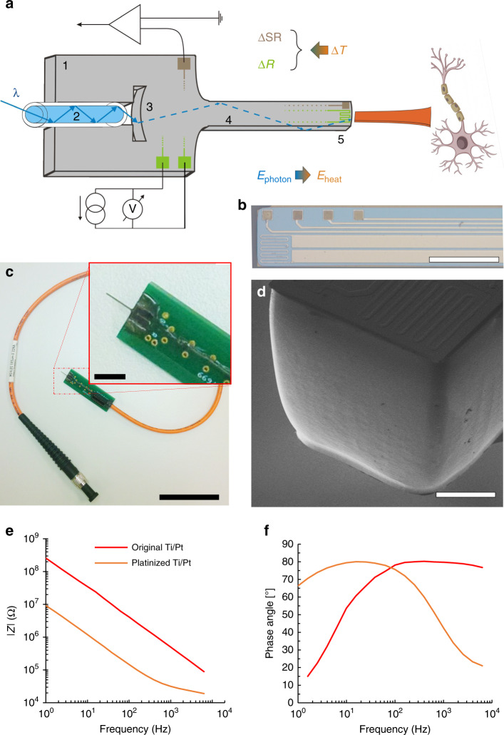
Fig. 1. A silicon neural microprobe designed for infrared stimualtion/inhibition of neuronal firing and interrogation of extrcellular potentials and temperature.
a Components of the implantable microsystem. 1: Si substrate, 2: multimode optical fiber, 3: cylindrical coupling lens, 4: shaft with multiple functionality, 5: probe tip. Figure is not to scale. b Optical microscopy image of the optrode tip. (Scale bar shows 300 µm) c Photo of an assembled optrode device. Scale bar of the picture with small and larger magnification are 3 cm and 5 mm, respectively. d Scanning electron micrograph representing the surface quality of the optrode tip. (Scale bar shows 100 µm) e, f Amplitude and phase diagrams of the impedance of the electrophysiological recording sites, respectively

