Fig. 2.
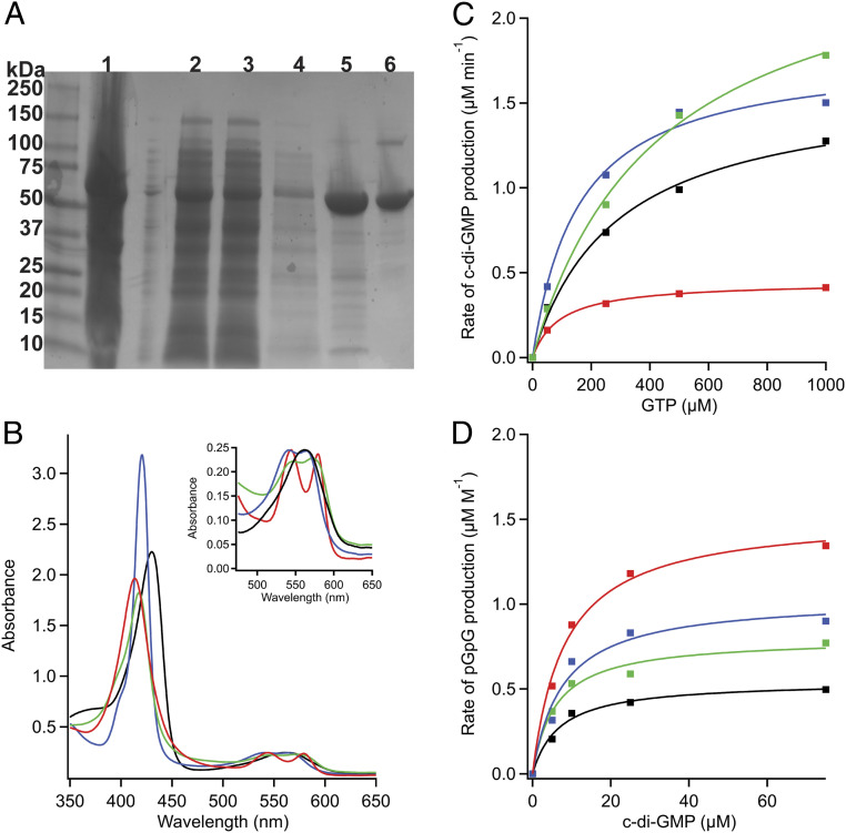
Biochemical characterization of DcpG. (A) SDS-PAGE. Lanes: 1, cell pellet; 2, supernatant; 3, flow through; 4, wash; 5, nickel column; 6, S200 gel filtration column. DcpG is recalcitrant to complete unfolding, yielding mainly monomer with a small dimer band on SDS-PAGE gels. (B) UV-visible spectra of DcpG. Fe(II), black; Fe(II)-O2, red; Fe(II)-CO, blue; Fe(II)-NO, green. The Soret band of DcpG is sensitive to the identity of the axial ligand. (Inset) The α/β bands exhibit increased splitting as the diatomic gases bind to the heme. (C) Representative Michaelis–Menten curves for DcpG Fe(II) (black), Fe(II)-O2 (red), Fe(II)-NO (green), and Fe(II)-CO (blue) DGC activity. (D) Representative Michaelis–Menten curves for DcpG Fe(II) (black), Fe(II)-O2 (red), Fe(II)-NO (green), and Fe(II)-CO (blue) PDE activity.

