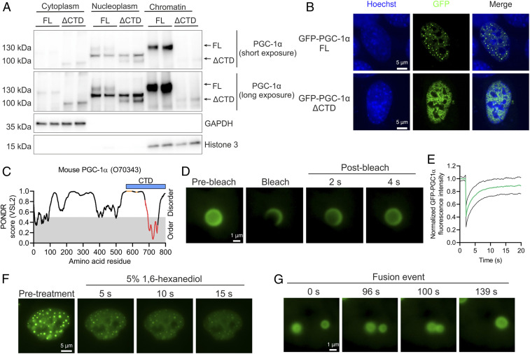
Fig. 4.
PGC-1α is localized within liquid-like nuclear condensates. (A) Subcellular fractionation of C2C12 myotubes expressing PGC-1α FL or ΔCTD. (B) Nuclear localization of GFP–PGC-1α FL or ΔCTD fusion proteins transfected in C2C12 myoblasts. (C) Predictor of Natural Disordered Regions (PONDR) analysis of mouse PGC-1α protein, with orange dots, red dots, and blue bar representing RS domains, RRM, and the CTD region under investigation, respectively. (D and E) Live-cell imaging (D) and quantification (E) of GFP–PGC-1α FL FRAP experiments in C2C12 myoblasts. Green and black lines denote mean and SD of FRAP quantification, respectively. (F) Live-cell imaging of 5% 1,6-hexanediol treatment of C2C12 myoblasts transfected with GFP–PGC-1α FL. (G) Live-cell imaging of a GFP–PGC-1α FL droplet fusion event in C2C12 myoblasts. Immunoblots and microscopy images are representative of at least three independent experiments each in triplicate.

