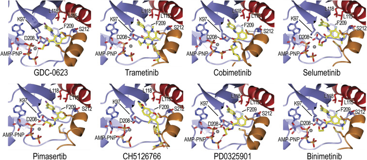
Fig. 2.
Details of MEK allosteric inhibitor binding. Inhibitors are shown in stick form with carbon atoms colored yellow. Hydrogen bonds are shown as dashed lines. Structural elements are colored as in Fig. 1 (A-loop helix orange, αC helix red), and selected side chains that form the allosteric binding pocket are labeled. Note that all inhibitors form a hydrogen bond with the mainchain amide of MEKS212 in the A-loop helix. Refer also to SI Appendix, Fig. S3 for corresponding electron density maps.

