Fig. 1.
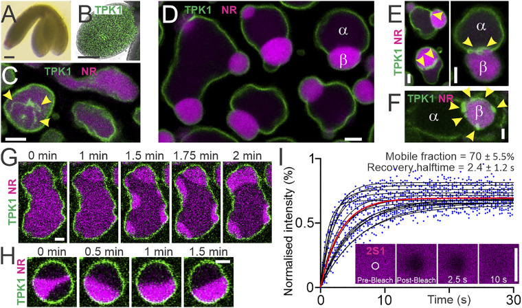
Liquid droplets wet and deform vacuolar membranes in living plant embryos. (A) A. thaliana embryo at walking stick developmental stage. (B) Embryonic cotyledon (leaf) expressing the tonoplast protein GFP-TPK1 (membrane, green). (C) Homogeneous vacuolar lumina characteristic of young vacuoles. Arrowheads, tonoplast-derived nanotubes. Individual frame from Movie S1. (D) Vacuolar liquid subcompartments and wet enclosing tonoplast. The droplet interface causes vacuole deformation and budding. (E and F) Tonoplast nanotubes wet the droplet interface. (G and H) Spontaneous droplet formation, flow, fusion, and repositioning observed by live-cell imaging. Snapshots from data shown in Movies S3 and S4. (I) Individual droplet FRAP data (blue dots). Fitted curves, black lines, n = 14 across three independent experiments. Red line, global fit. (Inset) Representative time series. Mean ± SD are shown. Confocal live-cell imaging. Vacuolar lumina (magenta) stained by 20 µM neutral red (NR) or expression of 2S1-GFP. (Scale bars: white, 2.5 µm; black, 100 µm.)

