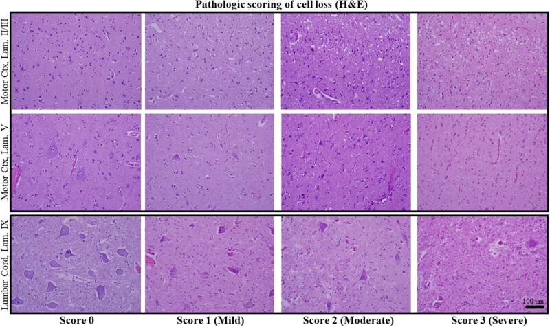
FIGURE 1.
Pathologic scoring of cell loss in H&E-stained autopsy material. Scores ranged from no evident cell loss (score 0; left-most column) to severe cell loss (score 3; right-most column) (see “Materials and Methods” for detail). The top 2 rows show lamina II and III (top row) and lamina V (middle row) in 4 ALS patients with scores 0 − 3. In the left-most panel of the middle row (score 0), large pyramidal cells (Betz cells) in lamina V are evident and in the score 3 example this layer is depleted of neurons. The bottom row shows residual α-motor neurons in the lateral portion of Lamina IX of the lumbar cord, ranging from score 0 (a non-ALS example; no ALS case in this study was score 0) to scores 1 − 3 (study samples shown). All images taken at ×200 magnification and the scale bar in the bottom right panel applies to all images.

