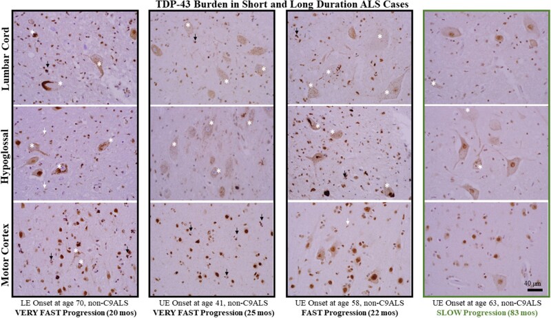
FIGURE 3.
TDP-43 burden in short and long duration ALS cases. TDP-43 burden in 3 short-lived, rapidly progressive patients (first 3 columns) and one long-lived, slowly progressive patient (right-most column). For each set of patient images, N-terminal TDP-43 staining is shown for lumbar cord (top row), hypoglossal nucleus (middle row) and motor cortex (bottom row). The images are representative of study samples and demonstrate greater TDP-43 inclusion pathology in the LMN pools of more rapidly progressive patients. The images also show the heterogeneity of neuronal pathology (white asterisks), including granular “pre-inclusions” with loss of nuclear staining (see text for detail), short neurites (white arrows), and oligodendroglial cytoplasmic inclusions (black arrows). Note that in several images the granular, pre-inclusion pathology with loss of nuclear staining is predominant (e.g. LMN pools of the patient shown in the second column with onset at age 41). All images taken at ×400 magnification and the scale bar in the bottom right panel applies to all images.

