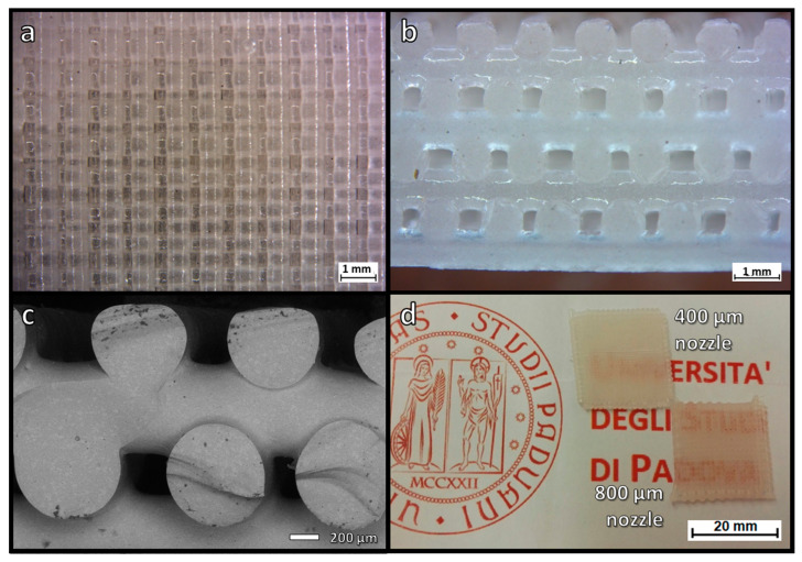
Figure 5.
Microstructural details of translucent scaffolds from LCD glass: (a,b) optical stereomicroscopy top and views of a scaffold from 800 µm nozzle (8 layers); (c) scanning electron micrograph of the fracture surface of a scaffold from 800 µm nozzle (4 layers); (d) visual comparison of ‘tiles’ from the overlapping of 4 layers.

