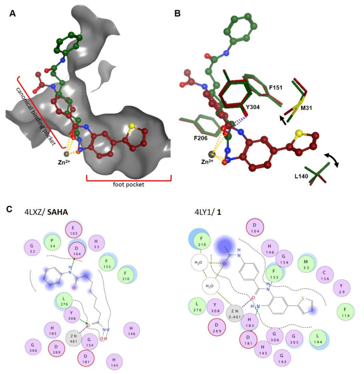
Figure 4.
Structural overlay of HDAC2 complexed with SAHA (PDB ID: 4LXZ) and the benzamide compound 1 (PDB ID: 4LY1; Lauffer et al. [30]). (A) Clipped binding pocket indicating the canonical binding pocket for substrate recognition and the widened foot pocket, which is also referred to as the acetate release channel. The zinc ion is shown as a gray sphere, and the clipped surface of the pocket is colored in gray. Metal bonds are shown as dotted orange lines. (B) L140 changes rotamers and M31 shifts to the side to open the foot pocket for the thiophenyl moiety. Hydrogen bonds are indicated as magenta dotted lines. (C) The 2D molecular interactions of SAHA and 1 within the canonical binding site of HDAC2. Side-chain hydrogen bonds are indicated by dotted green lines, whereas backbone hydrogen bonds are indicated as blue dotted lines. Metal complexation is shown using dotted magenta lines, solvent contacts are shown as dotted ochre lines, and exposure is illustrated with blue shading. Residues are numbered according to the crystal structure; for canonical numbering, refer to (A) and (B).

