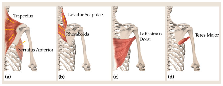
Figure 2.
Drawing showing the main lines of action of (a) the muscles trapezius (upper, middle, lower fibers), serratus anterior; (b) rhomboids and levator scapulae. Posterior view showing the location of the muscles latissumus dorsi (c) and teres major (d), constituting the posterior border of the axillary region.

