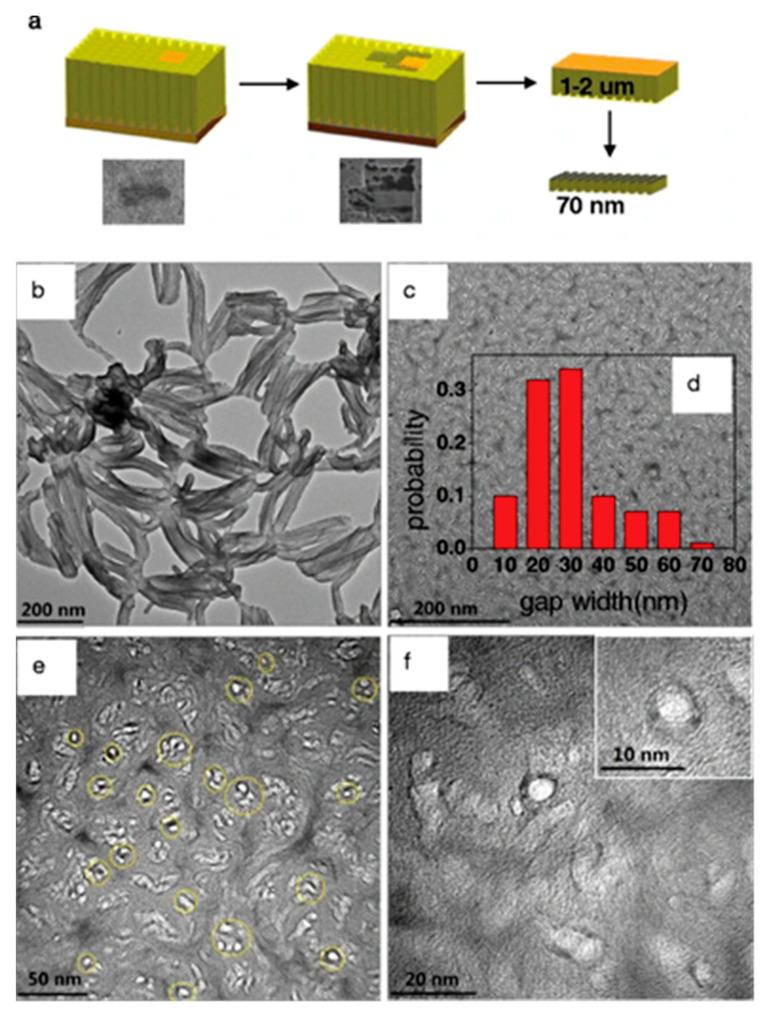
Figure 4.
(a) Focused ion beam (FIB) sample preparation (nanoindentation) and TEM micrographs; (b) cross-sectional TEM images of VACNT; (c,e,f) TEM images of the VACNTs/PANi nanocomposite membrane cross-section at different resolutions; and (d) histogram of the gap width distribution between the nanotubes (insert on (c)). Reproduced from Ding et al. (2015) [75] under the Creative Commons Licence BY 4.0. Springer Nature. Copyright © 2021, Ding et al.

