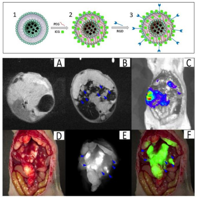
Figure 9.
Synthesis of SPIO@liposome-ICG-RGD (upper panel). (1) SPIO nanoparticles encapsulated in liposomes. (2) Magnetoliposome functionalized with ICG molecules. (3) Conjugation of RGDs peptide; orthotopic liver tumors with intrahepatic metastasis (lower panel). (A) The MRI image before SPIO@liposome-ICG-RGD injection. (B) The MRI signal was decreased in normal liver tissue (CNR: 14.6 ± 9.9) after targeting probe injection and the disseminated tumor nodes (0.9 ± 0.5 mm) (blue arrows) can be clearly defined. (C) Liver tumors confirmed by bioluminescence imaging. (D) Surgical guidance by intraoperative FMI-NIR. (E) The implanted liver tumor tissue (0.7 ± 0.3 mm) (blue arrows) exhibits obvious contrast (TBR: 2.3 ± 0.5) in colour and texture with normal liver tissues. (F) Merged colour and fluorescence image. Figure adapted from [94], Copyright 2017 Oncotarget.

