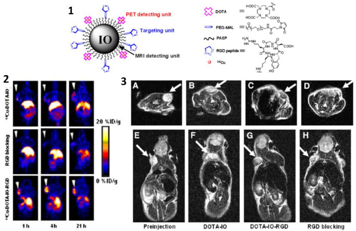
Figure 12.
(1) Illustration of PET/MRI probe based on iron oxide nanoparticle. (2) Decay-corrected whole-body coronal PET images of nude mouse bearing human U87MG tumor at 1, 4, and 21 h after injection of different PET/MRI probes based on iron oxide nanoparticles. (3) T2-weighted MR images of nude mice bearing U87MG tumor before injection of iron oxide nanoparticles (A,E) and at 4 h after tail-vein injection of DOTA-IO (B,F), DOTA-IO-RGD (C,G), and DOTA-IO-RGD with blocking dose of c(RGDyK) (D,H). Figure adapted from [103], Copyright 2008 Society of Nuclear Medicine, Inc.

