Figure 1.
ILC2s Proliferate in PcAT After MI
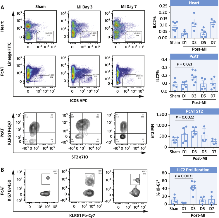
Flow cytometric analysis detects the expansion of ILC2 3 days post-MI (A) in heart (top) and pericardial adipose tissue accompanied with elevated ST2 surface expression (bottom), and increased proliferation (Ki67) (B). ILC2 proliferation in the PcAT was inhibited by injections of sST2, less pronounced in the heart (C). Representative images shown. Mann-Whitney U test. ILC2 = type 2 innate lymphoid cells; MI = myocardial infarction; PcAT = pericardial adipose tissue; sST2 = soluble ST2.


