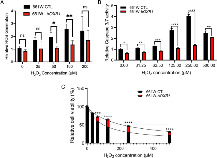
Figure 2.
Protective effects of hOXR1 in the 661W cone-derived photoreceptor cell line after H2O2 treatment. (A) ROS levels in 661W cells overexpressing hOXR1 normalized to control cells was determined using DCF-DA. Fluorescence was measured 20 minutes after treatment with the indicated concentrations of H2O2. (B) Caspase activity of 661W cells overexpressing hOXR1 (MVL120 cells) normalized to control (CTL) cells was determined with the Caspase 3/7-Gloassay 16 hours following the addition of the indicated concentrations of H2O2. (C) Cell titer-Gloassay was used to determine percent cell viability 16 hours after the addition of the indicated concentrations of H2O2. * P < 0.05, ** P < 0.01, *** P < 0.001, **** P < 0.0001, ns = nonsignificant, 2-way ANOVA test. Data are mean ± SD from two independent experiments, with three sets of samples per experiment.

