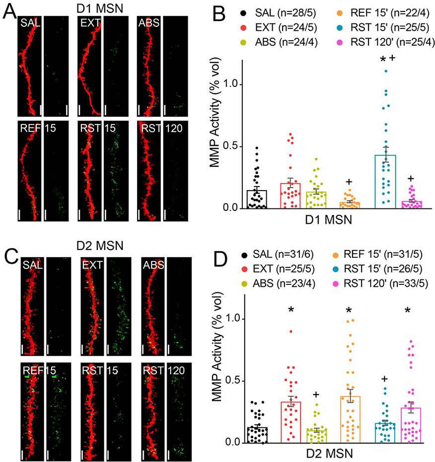
Figure 2. MMP-2 and MMP-9 activity localize differentially around D1- and D2-MSNs during heroin seeking and extinction.
A) Representative images of 3D reconstruction of mCherry-labeled D1 MSN dendritic segments (red) and MMP gelatinolytic puncta (green) localized within 300 nm of dendrite surface for each group yoked saline (SAL), withdrawn with extinction (EXT), withdrawn without extinction (ABS), 15 min in the extinguished environment (REF 15’), RST 15’ and RST 120’. Bar=7 μm. B) Transient heroin seeking induces significant increase in active MMP puncta localization around D1 MSNs (Nested ANOVA; F(4,17)=6.41; p=0.003). c) Representative images of 3D reconstruction of mCherry-labeled D2 MSN dendritic segments. Bar=7 μm. d) Extinction training induced increase in active MMP puncta localization around D2 MSNs (Nested ANOVA; F(5, 24)=6.50; p<0.001). Data are shown as mean ± SEM. N represents number of neurons quantified over number of in each condition. *p<0.05 compared to SAL, +p<0.05 compared to EXT, using Sidak post hoc test.

