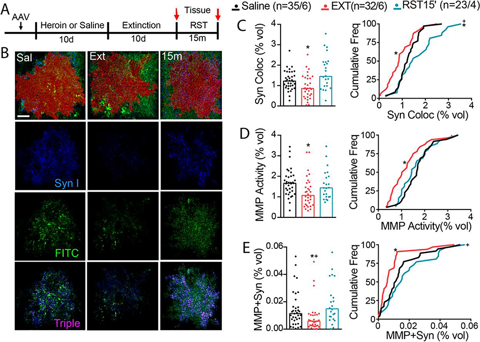
Figure 5. Heroin cues increase astroglia association with synaptic MMP-2,9 activity.
A) Experimental timeline outlining heroin self-administration, extinction, and reinstatement. Rats were trained to self-administer heroin and were subsequently extinguished to the cues associated with heroin infusions. B) Imaris quantification workflow for MMP-Synapsin I colocalization. Representative micrographs of digitized 3D model of mCherry-labeled astrocyte (red) for each treatment group (Sal, Ext, and RST 15 m). C) Synaptic contact between astrocyte membrane and presynaptic marker synapsin I was reduced after heroin extinction, while cued heroin seeking transiently restored peripheral astroglial processes to synapse (left panel shows raw data points, Kruskal-Wallis=13.32, p=0.001; right panel shows frequency plots, Kolmogorov-Smirnov=0.393, p=0.022). D) MMP activity co-registered with astrocyte membrane was decreased after heroin extinction (left panel shows raw data points, Kruskal-Wallis=10.06, p=0.007; right panel shows frequency plots, Kolmogorov-Smirnov=0.386, p=0.028). E) MMP gelatinolytic puncta and synapsin co-registry was reduced under extinguished conditions, while transiently increased during cue-induced heroin seeking (left panel shows raw data points, Kruskal-Wallis=12.80, p=0.002; right panel shows frequency plots, Kolmogorov-Smirnov=0.399, p=0.019 for SAL vs EXT, Kolmogorov-Smirnov=0.483, p=0.005 for EXT vs RST). N represents number of cells quantified over number of animals in each condition. Data are shown as median. *p<0.05 compared to SAL, +p<0.05 compared to RST using Dunn post hoc test.

